
Page 539 - IJB-9-5
P. 539
International Journal of Bioprinting GelMA/PEG-TA IPN networks for 3D bioprinting
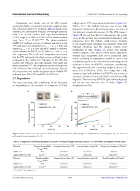
Compressive and tensile tests of the IPN showed collagenase at 37°C) were monitored in time (Figure 5A).
significantly higher compressive and tensile toughness than Within 25 h, the GelMA hydrogel was almost fully
that of the pure GelMA-UV hydrogel (Table 2). Based on the degraded. Compared to the initial specimen, the shape of
literature, the compressive modulus of hydrogels prepared this hydrogel already deformed at 6 h. The SEM image in
from 5 to 10 wt% GelMA with high functionalization Figure 5B showed that after 6 h degradation, the patchy
(>75%) range from 5 kPa to 20 kPa and the tensile modulus pores in the gel had been progressively degraded, with
range from 15 to 35 kPa [13,45,46] . The photo-crosslinked appearance from the initially small porous structure
GelMA-UV hydrogel was easy to deform compared to the to the macroporous structure. The integrity of the IPN
IPN and had a low compressive (E C-mod = 21 ± 6 kPa) and hydrogel remained, and the samples became more
tensile (E T-mod = 33 ± 6 kPa) modulus, similar to reported transparent in time (Figure 5B, insets). The initially
values, whereas the IPN E and E were 82 ± 2 and 111 ± 4 whitish samples, likely due to micro phase separation,
T
C
kPa, respectively. The tensile and compressive tests showed became fully transparent after 168 h incubation time,
that the IPN withstands both high stresses and high strains which according to degradation profile of GelMA has
compared to the GelMA-UV hydrogels. In the IPN, the
GelMA and 8PEGTA networks integrate both rigid and completely degraded. We infer that the remaining porous
5
elastic properties [35,47] . The compressive and tensile toughness structure is from the 8PEGTA enzymatic crosslinking.
5
of the IPN were 5.98 ± 0.56 and 2.93 ± 0.60 N/mm , whereas The approximately 30% remaining weight is close to the
2
the compressive and tensile toughness of the GelMA-UV mass ratio of 8PEGTA (25%). The change into a fully
5
hydrogels were 1.66 ± 0.21 and 0.48 ± 0.11 N/mm . transparent gel indicated that the 8PEGTA was present as
2
5
a second network and only the GelMA network was fully
3.7. Degradation degraded. The remaining 8PEGTA5 gel is slowly degraded
The mass remaining and morphology of the hydrogels and no gel was observed after 8 weeks (Figure S5,
on degradation in the presence of collagenase (2 U/mL Supplementary File).
A B
Figure 4. Stress-strain curves (wet state) of GelMA-UV and GelMA/8PEGTA -IPN hydrogels in compression (A) and elongation (B). The tests were
5
performed at room temperature (n = 5).
Table 2. Compressive and tensile properties of a photo‑crosslinked single network (GelMA‑UV) and double crosslinked network (IPN).
Network Compressive modulus Average fracture force (N) Average fracture strain (%) Compressive toughness (N/mm )
2
(E ) (kPa)
C‑mod
GelMA-UV 21±6 10.52±0.12 52.12±0.33 1.66±0.21
GelMA/8PEG-TA -IPN 82±2*** 28.63±0.80 64.73±0.52 5.98±0.56**
5
Network Tensile modulus Average fracture force (N) Average fracture strain (%) Tensile toughness (N/mm )
2
(E ) (kPa)
T‑mod
GelMA-UV 33±6 0.07±0.01 48.43±4.05 0.48±0.11
GelMA/8PEG-TA -IPN 111±4*** 0.24±0.01 71.25±5.14 2.93±0.60**
5
**P < 0.01, ***P < 0.001, n = 5
Volume 9 Issue 5 (2023) 531 https://doi.org/10.18063/ijb.750