
Page 238 - IJB-9-6
P. 238
International Journal of Bioprinting Bioprinting in diabetic foot disease
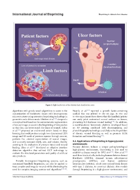
Figure 2. Eight key factors of the diabetic foot classification score
algorithms with greedy search algorithms to assist in the Nuutila et al. [119] reported a growth factor-containing
customization of biomimetic tissues with heterogeneous scaffold that was printed in situ on pigs. In vivo and
microstructures using extrusion bioprinting technology in in vitro experiments have shown that this handheld printer
geometric code-driven mode. Gholami et al. [115] designed a can easily print customized curved surfaces on tissues,
conceptual tool based on the semiautomatic segmentation promoting full-thickness wound healing [119] . In addition,
of wound images to assist in the bioprinting of skin patches a multifunctional hemostatic platform developed based
so that they can better match the shape of wounds. Zahia on 3D printing combined with electrospinning and
et al. [116] proposed an end-to-end system based on deep photolithography technology could help solve the problem
learning that could produce a single two-dimensional (2D) of chronic wound bleeding as well as promote ECM
image and 3D mesh of pressure injuries through sensors, formation and wound healing [120] .
perform fine external segmentation of wound images,
and accurately measure depth, area, and volume, thereby 4.2. Applications of bioprinting in hyperglycemic
assisting in the evaluation of pressure injury and wound environments
healing. Zhao et al. [117] developed an adaptive interface Because diabetes induces a unique pathophysiological
detection algorithm that utilized OCT technology to high-glucose environment, bioprinting is first used to
evaluate the structural parameters and quality of artificial establish a disease model for DFU skin [121] . Kim et al. [122]
skin products. used three kinds of cells, including human diabetes dermal
fibroblasts (dHDFs), diseased human subcutaneous
Portable biointegrated bioprinting systems, such as preadipocytes (dHPAs), and human epidermal
customized handheld bioprinters, can also be applied to keratinocytes (nHEKs), which were isolated from donors
treat complex and irregular wounds while eliminating the with type 2 diabetes, to construct diabetic skin models
need for complex imaging systems and algorithms [118-120] . through bioprinting in a high-glucose environment, and
Volume 9 Issue 6 (2023) 230 https://doi.org/10.36922/ijb.0142