
Page 478 - IJB-9-6
P. 478
International Journal of Bioprinting Bioprinting cell-laden protein-based hydrogel
the structure’s stability . The mechanism is thought to be (typically titanium or gold) is deposited on top of the
[80]
controlled via cell interactions and migrations, which is donor substrate. In response to the absorption of energy,
analyzed in some cases by means of phase field theory [83,84] . bioink droplets of defined sizes are formed. Although
Besides, cell aggregate maturation is a crucial step; in this technique is capable of printing materials with high
fact, changing the microenvironment in cell aggregates viscosities and high cell densities at extremely high
simulates tissue formation in vivo to accelerate maturation resolutions, it is limited by its excessive cost and inability
[25]
and generate stable, functional tissues . to print large constructs . In this regard, a number of TE
[85]
projects have been successfully performed using the multi-
In multi-cell bioprinting, subdivided into inkjet cell bioprinting method [91-98] . Compared to cell aggregate
bioprinting, extrusion-based bioprinting, and laser- and single-cell bioprinting, Table 1 summarizes the benefits
assisted bioprinting , cells are suspended in hydrogels and drawbacks of this method.
[86]
called bioinks . Employing this approach, cells are printed
[87]
randomly according to their concentration percentage in In summary, selecting the appropriate bioprinting
the bioink, and a scaffold is created afterward . Inkjet technique requires consideration of both the fabrication
[88]
printing is based on the deposition of bioink drops in a process and the cell requirements. Bioprinting of cell
predetermined way to create a final multi-layer pattern. aggregates is often necessary to produce large tissues
As a result of thermal or piezoelectric changes, pressure in size or have tight junctions between cells. The use of
pulses generate drops with a defined volume in the single-cell bioprinting permits researchers to deposit
range of picoliters. Commercial thermal printers utilize microenvironment components and stem cells in precisely
heating elements to expel ink droplets and form vapor defined locations to study cell–matrix interactions. In
bubbles that are heated to 300°C for a few microseconds. contrast, multi-cell bioprinting has proven to be the most
Printing on a substrate in the Z-direction is possible extensively used and advanced technique among all, for
with a micropositioning stage . Despite possessing a PBHs in particular.
[25]
relatively low printing resolution, this printing process
provides rapid scaffold production at an affordable cost. Of 3. Microenvironmental factors on bioprint-
note, low-viscosity materials and low cell concentrations ing the cell-laden PBHs
should be used to avoid nozzle clogging . Extrusion- In bioink formulation, it is crucial to consider the main
[89]
based bioprinters disperse bioinks as strands via a screw material and concentration as key parameters in order
plunger or an air pump; to be more specific, the dispenser to ensure the process’s reproducibility and enhance
is mounted on a robotic stage, enabling the printing head printability. Natural structural proteins, such as collagen,
to move in three directions. As a result of their design, elastin, silk fibroin, and fibrin, are particularly noteworthy
extrusion-based bioprinters can be used with hydrogels for their physiological and biological cues, contributing
having different viscosities and cell densities, and to the development of bioinks. Additionally, PBHs are
compared with inkjet-based bioprinting, there is less risk environmental friendly, renewable, and tend to exhibit
of clogging. However, if viscous hydrogels are utilized, a excellent biocompatibility, strength, elongation, toughness,
longer printing time and higher mechanical stresses may and slow degradability. All these remarkable materials’
reduce the viability of encapsulated cells by 40–80% . In characteristics originate from the proteins’ structure.
[90]
laser-based bioprinting, the bioink is transferred from one Indeed, features of proteins, such as hydrophobicity
substrate to another; to elucidate, pulses of the laser beam and bioactivity, which are the building units of living
are responsible for this transfer, and in order to control the organisms, depend on the amino acid constituents that are
transfer of energy, a thin layer of energy-absorbing material at the primary level, resulting in the folding of secondary
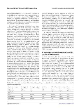
Tables 1. Benefits and drawbacks of different bioprinting techniques based on the format of the cells and the number of cells
produced during printing
Bioprinting technique Printing speed Printing throughput Resolution Cell viability Cell density Cost
Single-cell High Low High Medium Low High
Cell aggregate Medium High Low High High Medium
Multi-cell
Inkjet High Medium Medium Medium Low Low
Extrusion-based Low High Low Medium High Low
Laser-assisted Medium Medium High High Medium High
Volume 9 Issue 6 (2023) 470 https://doi.org/10.36922/ijb.1089