
Page 125 - v11i4
P. 125
International Journal of Bioprinting 3D bioprinting for translational toxicology
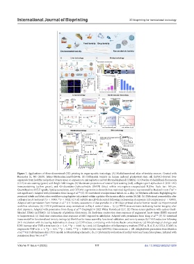
Figure 7. Applications of three-dimensional (3D) printing in organ-specific toxicology. (A) Multidimensional atlas of toxicity sources. Created with
Biorender [û, NP. (2025). https://BioRender.com/5bdwv4e. (B) Differential toxicity in human induced pluripotent stem cell (hiPSC)-derived liver
organoids from healthy and patient donors upon co-exposure to microplastics and tetrabromobisphenol (TBBPA). (a) Overlay of cholyl-lysyl-fluorescein
(CLF) in situ staining (green) with bright field images. (b) Maximum projections of neutral lipid staining (red), collagen type 1 alpha chain 1 (COL1A1)
immunostaining (yellow green), and 4’,6-diamidino-2-phenylindole (DAPI) (blue) within microsphere-encapsulated N_Dos. Scale bar: 100 μm.
Quantification of CLF uptake, lipid accumulation, and COL1A1 expression is shown below; statistical significance was assessed by Student’s t-test (“ns” =
not significant). Adapted with permission from Liang et al. (C) 3D convoluted renal proximal tubule-on-a-chip. (a) Nephron schematic highlighting the
189
proximal tubule and fabrication workflow using fugitive ink printed within a gelatin–fibrin extracellular matrix (ECM). (b) Diffusional permeability after
cyclosporine A treatment (*p < 0.003, **p < 0.02); (c) Cell viability in a 2D dish control following cyclosporine A exposure (all comparisons p < 0.005).
Adapted with permission from Homan et al. (D) Toxicity assessment of dust particles in a 3D inkjet-printed alveolar barrier model: (a) Experimental
19
workflow schematic; (b) CCK-8 proliferation assay normalized to Day 1 control (data = 1); (c) TEER measurements indicating barrier integrity after
dust exposure. Adapted with permission from Kang et al. Copyright © 2022 Wiley Periodicals LLC. (E) Tissue-sensor platform with carbon-black-
190
blended PDMS (cpPDMS): (a) Schematic of platform fabrication; (b) Real-time contraction dose-response of engineered heart tissue (EHT) exposed
to isoproterenol; (c) Real-time contraction dose-response of EHT exposed to nifedipine. Adapted with permission from Yong et al. (F) 3D intestinal
191
tissue model for functional and toxicity testing: (a) Workflow for tissue assembly, functional validation, and toxicity assays; (b) TEER reduction following
24 h incubation with increasing indomethacin doses; (c) LDH release correlating with indomethacin concentration; (d) Morphological changes and
LDH increase after TNF-α treatment (n = 5–6, ***p < 0.001 by t-test); (e) Upregulation of inflammatory markers (TNF-α, IL-6, IL-8, CCL2, ICAM) in
response to TNF-α (n = 3; **p < 0.01, ***p < 0.001, ****p < 0.0001 by two-way ANOVA). Data are mean ± SD. Adapted with permission from Madden
et al. (G) Full-thickness skin (FTS) model: (a) Bioprinting schematic; (b, c) Cytotoxicity evaluation of methyl violet and hexachlorophene. Adapted with
192
permission from Wei et al. 193
Volume 11 Issue 4 (2025) 117 doi: 10.36922/IJB025210209