
Page 126 - v11i4
P. 126
International Journal of Bioprinting 3D bioprinting for translational toxicology
For instance, Homan et al. employed an advanced 4.3. Lung toxicology
19
3D bioprinting technique to fabricate in vitro 3D human The lungs, as the primary site for gas exchange and
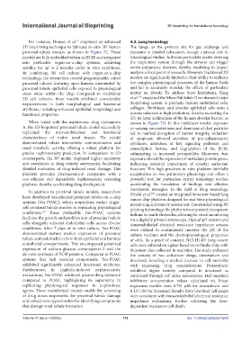
proximal tubule models, as shown in Figure 7C. These exposure to inhaled substances, occupy a pivotal role in
models are fully embedded within an ECM and integrated toxicological studies. Airborne particulate matter entering
into perfusable organ-on-a-chip systems, achieving the respiratory system through the airways can trigger
stability for up to 2 months under in vitro conditions. severe pulmonary diseases, thereby rendering its impact
By combining 3D cell culture with organ-on-a-chip analysis a focal point of research. However, traditional 2D
technology, the researchers created programmable, coiled models are significantly limited in their ability to replicate
proximal tubules featuring open lumens surrounded by the complex physiological processes of the human body
proximal tubule epithelial cells exposed to physiological and fail to accurately simulate the effects of particulate
shear stress within the chip. Compared to traditional matter on alveoli. To address these limitations, Kang
190
2D cell cultures, these models exhibited considerable et al. employed the MicroFab Jetlab II on-demand inkjet
improvements in both morphological and functional bioprinting system to precisely deposit endothelial cells,
attributes, including enhanced epithelial morphology and collagen, fibroblasts, and alveolar epithelial cells onto a
functional properties. porous substrate at high resolution, thereby recreating the
3D, tri-layer architecture of the human alveolar barrier, as
When tested with the nephrotoxic drug cyclosporin shown in Figure 7D. In this multilayer model, exposure
A, the 3D-bioprinted proximal tubule model successfully to varying concentrations and durations of dust particles
replicated the microarchitecture and functional led to marked disruption of barrier integrity, induction
characteristics of native renal tissues. The model of apoptosis, elevated secretion of pro-inflammatory
demonstrated robust intercellular communication and cytokines, activation of key signaling pathways and
renal metabolic activity, offering a robust platform for transcription factors, and degradation of the ECM,
precise nephrotoxicity prediction. In comparison to 2D culminating in increased permeability. Moreover, dust
counterparts, the 3D model displayed higher sensitivity exposure altered the expression of surfactant protein genes,
and correlation in drug toxicity assessments, facilitating indicating potential impairment of alveolar surfactant
detailed evaluation of drug-induced renal damage. This function. This high-precision in vitro platform faithfully
platform provides pharmaceutical companies with a recapitulates in vivo pulmonary physiology and offers a
cost-efficient and dependable nephrotoxicity screening powerful tool for particulate matter toxicology studies,
platform, thereby accelerating drug development. accelerating the translation of findings into effective
therapeutic strategies. In the field of drug toxicology,
In addition to proximal tubule models, researchers 200
have developed vascularized proximal tubule-on-a-chip Khalid et al. created an integrated biosensor-based lung
cancer chip platform designed for real-time physiological
systems (Vas-POAC), which outperform earlier single- monitoring and toxicity assessment. Constructed using 3D
cell proximal tubule models in replicating physiological printing technology, the platform incorporated transparent
conditions. These perfusable Vas-POAC systems indium tin oxide electrodes, allowing for visual monitoring
199
facilitate the growth and proliferation of proximal tubule via a digitally printed microscope. Optical pH sensors and
cells alongside adjacent endothelial cells across diverse transendothelial electrical resistance impedance sensors
conditions. After 7 days of in vitro culture, Vas-POAC were utilized to continuously monitor the pH of the
demonstrated mature marker expression of proximal culture medium and the electrophysiological properties
tubule and endothelial cells in both epithelial and luminal of cells. As a proof of concept, NCI-H1437 lung cancer
endothelial compartments. This encompassed polarized cells were cultured on a glass-based microfluidic chip, with
expression of sodium-glucose cotransporter-2 and the biosensor data collected in real time. The study evaluated
de novo synthesis of ECM proteins. Compared to POAC the toxicity of two anticancer drugs, doxorubicin and
systems that lack vascular components, Vas-POAC docetaxel, revealing a marked increase in cell mortality
exhibited significantly enhanced functional attributes. with increasing drug concentrations. Doxorubicin
Furthermore, in cisplatin-induced nephrotoxicity exhibited higher toxicity compared to docetaxel, as
evaluations, Vas-POAC exhibited greater drug tolerance confirmed through cell index assessments. Half-maximal
compared to POAC, highlighting its superiority in inhibitory concentration values calculated via linear
replicating physiological responses to nephrotoxic regression models were 6.791 µM for doxorubicin and
agents. These vascularized models enable the screening 0.137 nM for docetaxel. Results from live/dead cell assays
of drug doses responsible for proximal tubule damage were consistent with transendothelial electrical resistance
and unlock new opportunities for identifying compounds impedance evaluations, further validating the dose-
that disrupt renal tubule formation. dependent increase in cell death.
Volume 11 Issue 4 (2025) 118 doi: 10.36922/IJB025210209