
Page 121 - v11i4
P. 121
International Journal of Bioprinting 3D bioprinting for translational toxicology
3D porous architecture and an integrated heater design. Photopolymerization-based 3D printing, known for its
This innovation reduced response and recovery times high-resolution microstructural fabrication, has opened
to 214 and 222 s, respectively, and achieved a sensitivity new avenues in biosensing applications. Cao et al.
173
of 0.087% 1/ppm. Its dual-mode heating mechanism employed stereolithography to construct a paper-based
enhanced gas desorption efficiency, laying the groundwork microfluidic screen-printed electrode integrated with
for the development of wearable respiratory monitoring reduced graphene oxide-tetraethylene pentamine/Prussian
devices, as shown in Figure 6B. Expanding the frontier of blue composite materials, achieving a glucose detection
laser technology, Hecht et al. employed bubble-assisted limit of 25 μM, which is comparable to commercial
172
laser-induced forward transfer to achieve submicron-scale glucose meters. This integrated approach, which
patterning of polyimide-based bioinks on nitrocellulose combines microfluidic channels with electrochemical
substrates, as shown in Figure 6C. This approach enabled sensing units, significantly enhances detection efficiency
the fabrication of a multichannel C-reactive protein and supports personalized diabetes management.
detection system while maintaining material integrity Simultaneously, extrusion-based 3D printing has achieved
under high-energy laser exposure, thus offering a robust key breakthroughs in multifunctional sensor integration
manufacturing route for point-of-care diagnostic devices. due to its superior material compatibility. Marzo et al.
174
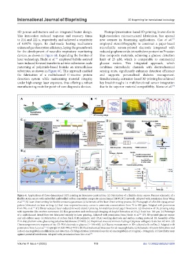
Figure 6. Applications of three-dimensional (3D) printing in biosensor construction. (A) Fabrication of a flexible strain sensor. Process schematic of a
flexible strain sensor with embedded multiwalled carbon nanotubes composite microchannel (MWCNT) network. Adapted with permission from Wang
170
et al. (B) Laser direct writing for flexible ammonia gas sensor. (a) Schematic of the laser direct writing process. (b) Photograph of a flexible zigzag sensor
pattern fabricated via laser writing. (c) Real-time response/recovery curves to ammonia concentrations from 75 to 400 ppm. Adapted with permission
171
from Wu et al. (C) Blister-actuated laser-induced forward transfer printing for multifunctional paper biosensors. (a) Overview of the printing setup
with typewriter-like receiver movement. (b) Key parameters and stroboscopic imaging of droplet formation at 2.5 μJ. Scale bar: 100 µm. (c) Prototype
of a multichannel lateral flow test fabricated entirely by laser printing. Adapted with permission from Hecht et al. (D) 3D-printed glucose sensor
172
and cell-culture assay. (a) Fabrication of carbon black (CB)-polylactic acid (PLA) working electrode and surface coating protocol. (b) Assembly of the
PLA chip platform onto glass using polydimethylsiloxane (PDMS). (c) Bioprinted sinusoid-mimetic hydrogel (alginate-collagen). Scale bar: 4.5 mm. (d)
Chronoamperometric response of the CB-PLA electrode to glucose (1–100 mM). (e) Glucose measurement in 3D-cultured cells on Day 2. Adapted with
175
permission from Lee et al. Copyright © 2023 Wiley-VCH. (E) Electrochemical biosensor for α2-macroglobulin. (a) Schematic of sensor fabrication and
anti-α2-macroglobulin modification and detection. (b) Magnetization hysteresis loops for α2-macroglobulin at 10 ng/mL–100 μg/mL. (c) Specificity assay
against potential interferents. Adapted with permission from Guo et al. 176
Volume 11 Issue 4 (2025) 113 doi: 10.36922/IJB025210209