
Page 119 - v11i4
P. 119
International Journal of Bioprinting 3D bioprinting for translational toxicology
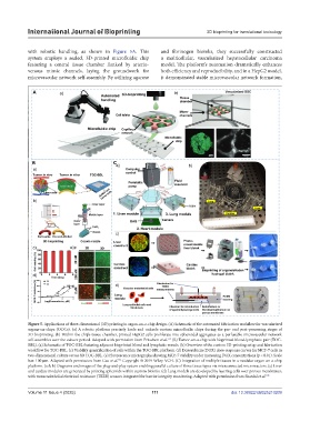
with robotic handling, as shown in Figure 5A. This and fibrinogen bioinks, they successfully constructed
system employs a sealed, 3D-printed microfluidic chip a multicellular, vascularized hepatocellular carcinoma
featuring a central tissue chamber flanked by arterio- model. The platform’s automation dramatically enhances
venous mimic channels, laying the groundwork for both efficiency and reproducibility, and in a HepG2 model,
microvascular network self-assembly. By utilizing agarose it demonstrated stable microvascular network formation,
Figure 5. Applications of three-dimensional (3D) printing in organ-on-a-chip design. (A) Schematic of the automated fabrication workflow for vascularized
organs-on-chips (OOCs). (a) A robotic platform precisely loads and unloads custom microfluidic chips during the pre- and post-processing stages of
3D bioprinting. (b) Within the chip’s tissue chamber, printed HepG2 cells proliferate into spheroidal aggregates as a perfusable microvascular network
159
self-assembles over the culture period. Adapted with permission from Fritschen et al. (B) Tumor-on-a-chip with bioprinted blood-lymphatic pair (TOC-
BBL). (a) Schematic of TOC-BBL featuring adjacent bioprinted blood and lymphatic vessels. (b) Overview of the custom 3D-printing setup and fabrication
workflow for TOC-BBL. (c) Viability quantification of cells within the TOC-BBL platform. (d) Doxorubicin (DOX) dose-response curves for MCF-7 cells in
two-dimensional culture versus 3D TOC-BBL. (e) Fluorescence micrographs showing MCF-7 viability under increasing DOX concentrations (p < 0.01). Scale
162
bar: 100 µm. Adapted with permission from Cao et al. Copyright © 2019 Wiley-VCH. (C) Integration of multiple tissues in a modular organ-on-a-chip
platform. (a & b) Diagrams and images of the plug-and-play system enabling parallel culture of three tissue types via interconnected microreactors. (c) Liver
and cardiac modules are generated by printing spheroids within custom bioinks. (d) Lung models are developed by layering cells over porous membranes,
with transendothelial electrical resistance (TEER) sensors integrated for barrier integrity monitoring. Adapted with permission from Skardal et al. 165
Volume 11 Issue 4 (2025) 111 doi: 10.36922/IJB025210209