
Page 205 - v11i4
P. 205
International Journal of Bioprinting Bioprinted osteoarthritis scaffolds
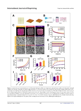
Figure 2. Construction and characterization of a 3D-bioprinted hydrogel scaffold. (A) Single-layer and multi-layer models of 3D-bioprinted hydrogel
scaffolds. (B) Schematic illustration of natural bioink preparation. (C) Structural profiles of the printed Gel–Alg hydrogel scaffolds; SEM images of non-
crosslinked and crosslinked Gel–Alg hydrogel scaffolds. (D and E) The viscosity (D) and modulus (E) of the natural bioink. (F) Stress–strain curves of
the Gel–Alg hydrogel scaffolds. (G) Statistical Young’s modulus of the Gel–Alg scaffolds. (H and I) Swelling performance (H) and water content (I) of the
Gel–Alg hydrogel scaffolds. (J) In vitro degradation performance of the scaffolds. (K) Porosity of the Gel–Alg hydrogel scaffolds. All statistical data are
represented as mean ± standard deviation (n = 3; *p < 0.05, **p < 0.01, ***p < 0.001). Scale bar: 1 mm (C). Abbreviations: ACs, articular chondrocytes; Alg,
alginate; GAGs, glycosaminoglycans; Gel, gelatin; SEM, scanning electron microscopy; sGAGs, sulfated glycosaminoglycans.
Volume 11 Issue 4 (2025) 197 doi: 10.36922/IJB025150136