
Page 207 - v11i4
P. 207
International Journal of Bioprinting Bioprinted osteoarthritis scaffolds
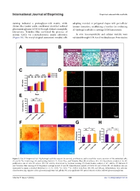
staining indicated a proteoglycan-rich matrix, while adopting rounded or polygonal shapes with pericellular
Alcian Blue (under acidic conditions) identified sulfated lacunae formation, establishing a baseline for evaluating
glycosaminoglycans (sGAGs) through distinct cyanophilic 3D hydrogel scaffolds in cartilage ECM homeostasis.
interactions. Toluidine Blue confirmed the presence of
anionic GAGs via γ-metachromatic purple coloration In vitro biocompatibility and cellular viability were
(Figure 3B). The morphological assessment revealed cells validated through CCK-8 and live/dead assays. Prior studies
Figure 3. The 3D-bioprinted Gel–Alg hydrogel scaffolds support the survival, proliferation, and extracellular matrix secretion of the embedded cells.
(A and B) The morphology (A) and staining (Safranin O, Alcian Blue, and Toluidine Blue) (B) of primary ACs. (C) Quantitative analysis of the AC
proliferation rate in vitro 2D culture. (D) Cell viability determined by live/dead staining. (E) Quantification analysis of AC viability. (F) Safranin O
and Toluidine Blue staining of 3D-bioprinted cartilage model. (G and H) Quantification analysis of GAGs (G) and sGAGs (H). All statistical data are
represented as mean ± standard deviation (n = 5; *p < 0.05, **p < 0.01, ***p < 0.001). Scale bars: 100, 200, and 500 µm. Abbreviations: ACs, articular
chondrocytes; Alg, alginate; GAGs, glycosaminoglycans; Gel, gelatin; NS, non-significant; OD, optical density; sGAGs, sulfated glycosaminoglycans.
Volume 11 Issue 4 (2025) 199 doi: 10.36922/IJB025150136