
Page 212 - v11i4
P. 212
International Journal of Bioprinting Bioprinted osteoarthritis scaffolds
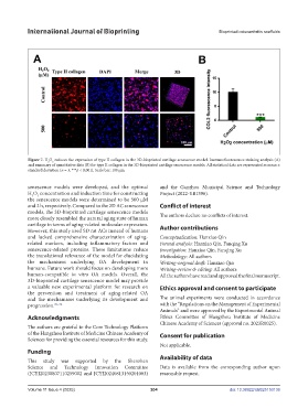
Figure 7. H O reduces the expression of type II collagen in the 3D-bioprinted cartilage senescence model. Immunofluorescence staining analysis (A)
2
2
and summary of quantitative data (B) for type II collagen in the 3D-bioprinted cartilage senescence models. All statistical data are represented as mean ±
standard deviation (n = 3; ***p < 0.001). Scale bar: 100 µm.
senescence models were developed, and the optimal and the Ganzhou Municipal Science and Technology
H O concentration and induction time for constructing Project (2022-YB1396).
2
2
the senescence models were determined to be 500 μM
and 2 h, respectively. Compared to the 2D AC senescence Conflict of interest
models, the 3D-bioprinted cartilage senescence models The authors declare no conflicts of interest.
more closely resembled the natural aging state of human
cartilage in terms of aging-related molecular expression. Author contributions
However, this study used SD rat ACs instead of humans
and lacked comprehensive characterization of aging- Conceptualization: Hanxiao Qin
related markers, including inflammatory factors and Formal analysis: Hanxiao Qin, Fanqing Xu
senescence-related proteins. These limitations reduce Investigation: Hanxiao Qin, Fanqing Xu
the translational relevance of the model for elucidating Methodology: All authors
the mechanisms underlying OA development in Writing–original draft: Hanxiao Qin
humans. Future work should focus on developing more Writing–review & editing: All authors
human-compatible in vitro OA models. Overall, the All the authors have read and approved the final manuscript.
3D-bioprinted cartilage senescence model may provide
a valuable new experimental platform for research on Ethics approval and consent to participate
the prevention and treatment of aging-related OA
and the mechanisms underlying its development and The animal experiments were conducted in accordance
progression. 70–72 with the “Regulations on the Management of Experimental
Animals” and were approved by the Experimental Animal
Acknowledgments Ethics Committee of Hangzhou Institute of Medicine
Chinese Academy of Sciences (approval no. 2023R0025).
The authors are grateful to the Core Technology Platform
of the Hangzhou Institute of Medicine Chinese Academy of Consent for publication
Sciences for providing the essential resources for this study.
Not applicable.
Funding
This study was supported by the Shenzhen Availability of data
Science and Technology Innovation Committee Data is available from the corresponding author upon
(JCYJ20230807110259002 and JCYJ20240813150201003) reasonable request.
Volume 11 Issue 4 (2025) 204 doi: 10.36922/IJB025150136