
Page 211 - v11i4
P. 211
International Journal of Bioprinting Bioprinted osteoarthritis scaffolds
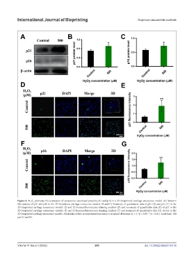
Figure 6. H O promotes the expression of senescence-associated proteins p21 and p16 in a 3D-bioprinted cartilage senescence model. (A) Western
2
2
blot analysis of p21 and p16 in the 3D-bioprinted cartilage senescence models. (B and C) Summary of quantitative data of p21 (B) and p16 (C) in the
3D-bioprinted cartilage senescence models. (D and E) Immunofluorescence staining analysis (D) and summary of quantitative data (E) of p21 in the
3D-bioprinted cartilage senescence models. (F and G) Immunofluorescence staining analysis (F) and summary of quantitative data (G) of p16 in the
3D-bioprinted cartilage senescence models. All statistical data are represented as mean ± standard deviation (n = 3; *p < 0.05, **p < 0.01). Scale bars: 100
µm (D and F).
Volume 11 Issue 4 (2025) 203 doi: 10.36922/IJB025150136