
Page 26 - v11i4
P. 26
International Journal of Bioprinting 3D-printed scaffolds for osteochondral defect
quality of the newly formed meniscal tissue was assessed lap shear test requires extra clamping or adhesive bonding
in vivo by the rabbit model. Compared to scaffolds without of the scaffold, which may introduce additional artifacts
MSC loading, the incorporation of MSCs into the scaffold in evaluation. While the peel test provides insight into
36
significantly improved scaffold integration, proteoglycan interface strength, its loading condition failed to replicate
synthesis, and the mechanical properties of the newly in vivo scenarios. Given these considerations, the interface
formed tissue. Similarly, Chung et al. demonstrated that shear test is generally regarded as the most appropriate
144
hUCMSCs-laden 4% HA hydrogel significantly enhanced method for evaluating interface strength. Additionally,
136
cartilage quality histologically, achieving a remarkable histological studies can reveal the tissue growth pattern,
collagen pattern and cell arrangement that closely resemble including cell density, cell alignment, and ECM, which
the structure of adjacent native cartilage. can serve as an important reference for assessing
interface integration. 139
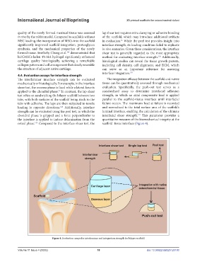
4.4. Evaluation assays for interface strength
The interlaminar interface strength can be evaluated The integration efficacy between the scaffold and native
mechanically or histologically. For example, in the interface tissue can be quantitatively assessed through mechanical
shear test, the osseous phase is fixed while a lateral force is evaluation. Specifically, the push-out test serves as a
applied to the chondral phase. In contrast, the lap shear standardized assay to determine interfacial adhesion
36
test relies on sandwiching the bilayer scaffold between two strength, in which an axial compressive load is applied
tabs, with both surfaces of the scaffold being stuck to the parallel to the scaffold–tissue interface until interfacial
tabs with adhesive. The laps are then subjected to tensile failure occurs. The maximum load at failure is recorded
loading in opposite directions. Additionally, interface and normalized to the total surface area of the scaffold’s
145
strength can be evaluated using the peel test, in which the luminal interface, enabling the calculation of the ultimate
141
chondral phase is gripped and a force perpendicular to interfacial shear strength. This parameter provides a
the interface is applied to induce delamination from the quantitative measure of the biomechanical integrity at the
osteal phase. Compared to the interface shear test, the scaffold–tissue interface (Figure 4).
146
Figure 4. Evaluation assays for interlaminar and integration strength for bilayer scaffold.
Volume 11 Issue 4 (2025) 18 doi: 10.36922/IJB025120100