
Page 294 - v11i4
P. 294
International Journal of Bioprinting Dual tuning of 3D-printed SilMA hydrogel
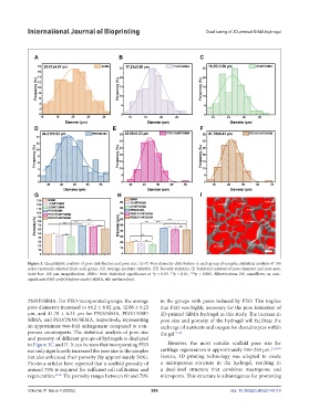
Figure 3. Quantitative analysis of pore distribution and pore size. (A–F) Pore diameter distribution in each group of samples, statistical analysis of 100
pores randomly selected from each group. (G) Average aperture statistics. (H) Porosity statistics. (I) Statistical method of pore diameter and pore area.
Scale bar: 100 µm; magnification: 1000×. Note: Statistical significance at *p < 0.05, **p < 0.01, ***p < 0.001. Abbreviations: NF, nanofibers; ns, non-
significant; PEO, poly(ethylene oxide); SilMA, silk methacryloyl.
2%NF/SilMA. For PEO-incorporated groups, the average in the groups with pores induced by PEO. This implies
pore diameters increased to 44.2 ± 6.02 µm, 42.06 ± 6.23 that PEO was highly necessary for the pore formation of
µm, and 41.78 ± 6.23 µm for PEO/SilMA, PEO/1%NF/ 3D-printed SilMA hydrogel in this study. The increase in
SilMA, and PEO/2%NF/SilMA, respectively, representing pore size and porosity of the hydrogel will facilitate the
an approximate two-fold enlargement compared to non- exchange of nutrients and oxygen for chondrocytes within
porous counterparts. The statistical analysis of pore size the gel. 37,38
and porosity of different groups of hydrogels is displayed
in Figure 3G and H. It can be seen that incorporating PEO However, the most suitable scaffold pore size for
not only significantly increased the pore size in the samples cartilage regeneration is approximately 150–250 µm. 21,39,40
but also enhanced their porosity (by approximately 50%). Herein, 3D printing technology was adopted to create
Previous articles have reported that a scaffold porosity of a microporous structure in the hydrogel, resulting in
around 70% is required for sufficient cell infiltration and a dual-level structure that combines macropores and
regeneration. 35,36 The porosity ranges between 60 and 70% micropores. This structure is advantageous for promoting
Volume 11 Issue 4 (2025) 286 doi: 10.36922/IJB025140118