
Page 344 - v11i4
P. 344
International Journal of Bioprinting GradGelMA 3D-bioprinted vascular skin
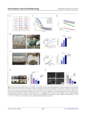
Figure 3. Characterization of gelatin methacryloyl (GelMA). (A) Quantification of the degree of functionalization of GelMA using proton nuclear magnetic
resonance spectroscopy. (B) Compression testing of samples, experimental procedures, and quantification of compression modulus. Scale bars: 5 and 10
mm. (C) Tensile testing of samples, experimental procedures, and quantification of tensile modulus. Scale bars: 5 and 10 mm. (D) Samples of GelMA before
and after freeze-drying, and quantification of swelling ratio. Scale bar: 10 mm. (E) Scanning electron microscopy images of GelMA: representative electron
micrographs and quantification of GelMA porosity. Scale bar: 100 µm, magnification 500×. (F) Determination of gelation point in GelMA hydrogels.
(G) Rheological characterization of GelMA. Data were analyzed via a one-way analysis of variance and are shown as mean ± standard deviation (*p < 0.05,
**p < 0.01, ***p < 0.001, n = 3). For a better overview, only the significant differences are indicated.
Volume 11 Issue 4 (2025) 336 doi: 10.36922/IJB025090069