
Page 348 - v11i4
P. 348
International Journal of Bioprinting GradGelMA 3D-bioprinted vascular skin
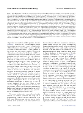
Figure 5. Skin cell-compatible composite ink. (A) Live/dead staining to assess the viability and stretching of human foreskin fibroblast (HFF) cells in
5%, 10%, 15%, and 20% (w/v) gelatin methacryloyl (GelMA). Scale bar: 300 µm; magnification: 50×. (B) F-actin/4ʹ,6-diamidino-2-phenylindole (DAPI)
staining of HFF cells cultured in 5% (w/v) GelMA for 10 days. Scale bar: 100 µm. (C) Cell counting kit-8 (CCK-8) assay to measure the proliferation of
HFF cells in 5%, 10%, 15%, and 20% (w/v) GelMA from Day 1 to Day 7. (D) Western blot analysis of collagen I protein expression in HFF cells cultured in
5%, 10%, 15%, and 20% (w/v) GelMA, quantifying collagen I expression using ImageJ. (E) Proliferation of human immortalized epidermal cells (HaCaT)
cells on the surface of 5%, 10%, 15%, and 20% (w/v) GelMA, with quantification of HaCaT cells surface coverage using ImageJ. Scale bar: 500 µm. (F)
Hematoxylin and eosin staining of HaCaT cells cultured on 20% (w/v) GelMA and collagen surfaces for 10 and 28 days, with quantification of epidermal
thickness using ImageJ. Scale bar: 100 µm; magnification: 50×. (G) Immunofluorescence staining of Involucrin (IVL) and Cytokeratin 14 (CK14) in
HaCaT cells cultured on 20% (w/v) GelMA and collagen surfaces for 10 and 28 days, with quantification of keratinocyte layer number and IVL/CK14
expression using ImageJ. Scale bar: 100 µm. (H) Fluorescence microscopy images of human umbilical vein endothelial cells (HUVECs) spreading in 3%
(w/v) GelMA at D1, D7, and D14, with quantification of junction, mesh, and branch formation using ImageJ. Scale bars: 200, 500 µm; magnification:
50×. (I) Immunofluorescence staining of platelet endothelial cell adhesion molecule-1 (PECAM-1/CD31) and F-actin in HUVECs cultured in 3% (w/v)
GelMA at Day 7 and Day 14, quantifying CD31 and F-actin expression using ImageJ. Scale bar: 100 µm. (J) Schematic diagram of the HUVECs migration
experiment from 3% (w/v) GelMA to 3% (w/v) GelMA or 5% (w/v) GelMA, with microscopy images of cell migration at D7 and D14, and quantification
of migration distance using ImageJ. Scale bar: 200 µm; magnification: 50×. The red dashed lines mark areas where partial cell extension and interconnected
microvascular structures are observed. Data were analyzed via a one-way analysis of variance and are shown as mean ± standard deviation (*p < 0.05, **p
< 0.01, ***p < 0.001, n = 3). For a better overview, only the significant differences are indicated. Abbreviations: ALI, air–liquid interface; FIB, fibrinogen;
GAPDH, glyceraldehyde-3-phosphate dehydrogenase; IOD, integrated optical density
displayed a more uniform and flat epidermal structure there are arterioles and venules. The arterioles and venules,
(Figure 5F). Involucrin (IVL) is a highly reactive soluble formed by their branching, pass through the dermis and
transamidase substrate protein present in keratinocytes form a rich microvascular network in the upper layer of
of the epidermis and other stratified squamous epithelia, the dermis (papillary layer), which supplies oxygen and
synthesized in the spinous layer. It is highly expressed in nutrients to the epidermis. 43,44 The construction of the
the granular layer and serves as a marker of the terminal microvascular network in the upper layer of the dermis
differentiation of keratinocytes. Cytokeratin 14 (CK14) is is of great significance for the construction of the skin
a member of the intermediate fiber albumen type I keratin epidermal barrier function. As shown in Figure S1,
family that pairs with type II keratin CK5 to form the basic Supporting Information, HUVECs were cultured in
keratin of stratified squamous epithelial keratinocytes, 3% (w/v), 5% (w/v), and 7% (w/v) GelMA hydrogels,
including the epidermis and non-keratinized stratified respectively, and observed under a microscope every
squamous epithelial mucosa. It is highly expressed in the 3 days for imaging. In the 3% (w/v) GelMA hydrogel
undifferentiated basal cell layer, containing stem cells, group, HUVECs began to extend and elongate by Day
and down-regulated in differentiated basal cell layers. The 3, and extensive microvascular formation was observed
GelMA and collagen groups had a cell structure of more by Day 9. In the 5% (w/v) GelMA hydrogel group, slight
than 10 layers, with no obvious difference between them, cell elongation was noted by Day 9, while no significant
similar to the structure of the human skin epidermis. changes were observed in the 7% (w/v) GelMA hydrogel
CK14 and IVL were significantly expressed in the GelMA group. In conclusion, the 3% (w/v) GelMA hydrogel is more
and fibrinogen groups (Figure 5G). The observed pattern suitable for HUVEC culture. When the 3% (w/v) GelMA
of CK14 and IVL co-expression in the same region may bio-ink containing HUVEC-green fluorescent protein
be attributed to the unique microenvironment of in vitro (1× 10 /mL) was cultured for 7 days after solidification,
6
culture, where the differentiation process of keratinocytes the vascular endothelial cells began to spread and intersect
might not fully replicate the in vivo scenario, leading to form some connection points, showing a tube-forming
to overlapping expression of markers that are typically phenomenon. On the 14th day, the tube-forming effect
spatially separated. The 20% (w/v) GelMA is more of vascular endothelial cells was apparent, producing
conducive to the growth and proliferation of HaCaT cells many branches, connection points, and grid structures
on its surface, and the keratinocytes on its surface have a (Figure 5H). PECAM-1/CD31 is a transmembrane
high degree of functional expression. Therefore, 20% (w/v) glycoprotein belonging to the immunoglobulin superfamily.
GelMA loaded with HaCaT cells was selected as the bio- It is a major marker of vascular endothelial cells. It is often
ink for constructing the epidermal base layer. used to identify the structure of vascular endothelial
The human vascular system is a complex and precise cells, and its expression level can reflect the functionality
network that spreads throughout the body and is of vascular endothelial cells. During the incubation
45
responsible for transporting oxygen and nutrients to all of 3% (w/v) GelMA bio-ink containing HUVEC-green
body parts while removing waste and carbon dioxide fluorescent protein (1 × 10 /mL) from 7 to 14 days, the
6
through blood circulation. In the subcutaneous tissue, expression of CD31 in vascular endothelial cells increased
Volume 11 Issue 4 (2025) 340 doi: 10.36922/IJB025090069