
Page 45 - v11i4
P. 45
International Journal of Bioprinting 3D bioprinting of nerve guidance conduits
conduits from laboratory research to commercialization model in 1881; however, the results were unsatisfactory.
37
and clinical applications and outlines potential future In 1982, Lundborg et al. attempted to bridge a 6 mm
38
research directions and development trends. nerve defect in rats with a hollow and clear silicone
conduit, demonstrating that axonal regeneration begins
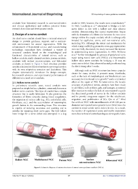
2. Design of a nerve conduit with the formation of a fibrin clot between the two nerve
An ideal nerve conduit should have a rational structural stumps within the empty conduit, which is subsequently
design to provide guidance, support, and a nutrient- invaded by capillaries, axons, and non-neuronal cells,
rich environment for axonal regeneration. With the including SCs. Although the extent and mechanisms by
advancement of biomaterial science and manufacturing which stump scaffolding promotes cross-gap regeneration
technology, researchers have developed a variety of were not fully elucidated, the study increased the interest
structural conduits based on the morphological and in understanding nerve regeneration. In 1983, Williams
39
functional characteristics of natural nerves, such as et al. further investigated the process of peripheral nerve
hollow conduits, multi-channel conduits, porous conduits, repair using neural stem cells (NSCs) and impermeable
conduits with surface microstructures, and bifurcated hollow silica nerve conduits by bridging a 10 mm rat
conduits, as shown in Figure 4. Each structure provides sciatic nerve defect. They observed axonal growth reaching
specific solutions for different barriers to nerve regeneration the distal stump after 3 weeks.
with its own adaptation scenarios and limitations. This Although such an NGC structure has been a popular
chapter systematically introduces the design concepts, choice for many studies, it presents many drawbacks,
key research advances, and experimental performances of such as the lack of morphological and biochemical cues
different structural nerve conduits. necessary for directional nerve growth, and a very limited
40
2.1. Hollow nerve conduit effect on the repair of PNI. To better mimic the bundle
In preliminary scientific cases, neural conduits were structure of peripheral nerves, attempts have been made
prepared as simple hollow cylinders, commonly known as to add fillers, such as fibers, gels, and sponges, as internal
hollow nerve conduits. This type of conduit has a simple filler matrices to reduce the lack of cues required to guide
structure that is easily fabricated. It also promotes the the directional growth of nerves in the hollow conduits
formation of fibrin networks during blood coagulation, and to provide exogenous support for the attachment,
41
the migration of various cells (e.g., SCs, endothelial cells, migration, and proliferation of SCs. Qin et al. filled
fibroblasts, etc.), and the accumulation of neurotrophic the artificial NGC with microfilaments of 80–120 μm in
growth factors in the surrounding tissue. This structure diameter and injected nerve growth factor (NGF) into the
is capable of reducing neuromas and scarring and is conduit to repair sciatic nerve defects in rats. As a result,
suitable for repairing short nerve gaps. The first hollow the number of regenerated axons and myelin maturation
bone bridge for a nerve defect was attempted in a dog were close to those of control autologous nerve grafts. Later,
Figure 4. Schematic diagram of various structures of current nerve guidance conduits (NGCs).
Volume 11 Issue 4 (2025) 37 doi: 10.36922/IJB025140120