
Page 42 - v11i4
P. 42
International Journal of Bioprinting 3D bioprinting of nerve guidance conduits
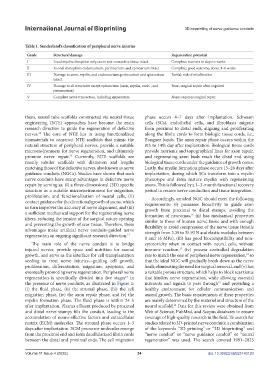
Table 1. Sunderland’s classification of peripheral nerve injuries
Grade Structural damage Regeneration potential
I Local myelin disruption only; axon and connective tissue intact Complete; recovers in days to weeks
II Axonal disruption; endoneurium, perineurium, and epineurium intact Complete; good outcome, about 4–6 weeks
III Damage to axon, myelin, and endoneurium; perineurium and epineurium Partial; risk of misdirection
intact.
IV Damage to all structures except epineurium (axon, myelin, endo-, and Poor; surgical repair often required
perineurium)
V Complete nerve transection, including epineurium None; requires surgical repair
them, neural tube scaffolds constructed via neural tissue phase occurs 4–7 days after implantation. Schwann
engineering (NTE) approaches have become the main cells (SCs), endothelial cells, and fibroblasts migrate
research direction to guide the regeneration of defective from proximal to distal ends, aligning and proliferating
nerves. The core of NTE lies in using functionalized along the fibrin cords to form biologic tissue cords, i.e.,
12
biomaterials to construct NTE scaffolds that mimic the Bungner bands. The axon repair phase occurs within the
natural structure of peripheral nerves, provide a suitable 8th to 14th day after implantation. Biological tissue cords
microenvironment for nerve regeneration, and ultimately provide nutrients and topographical lines for axon repair,
promote nerve repair. Currently, NTE scaffolds are and regenerating axon buds reach the distal end using
13
mostly tubular scaffolds with diameters and lengths biological tissue cords under the guidance of growth cones.
matching those of the defective nerves, also known as nerve Lastly, the myelin formation phase occurs 15–28 days after
guidance conduits (NGCs). Studies have shown that such implantation, during which SCs transform into a myelin
nerve conduits have many advantages in defective nerve phenotype and form mature myelin with regenerating
repair by serving as: (i) a three-dimensional (3D) specific axons. This is followed by a 1–3-month functional recovery
structure as a suitable microenvironment for migration, period to restore nerve conduction and tissue integration.
proliferation, and functionalization of neural cells, (ii) Accordingly, an ideal NGC should meet the following
contact guidance for the directional growth of axons, which requirements: (i) possesses bioactivity to guide axon
in turn improves the accuracy of nerve alignment, and (iii) growth from proximal to distal stumps, avoiding the
a sufficient mechanical support for the regenerating nerve formation of neuromas, (ii) has mechanical properties
16
fibers, reducing the tension of the surgical suture opening similar to those of human nerve tissue and with enough
and preventing the growth of scar tissue. Therefore, these flexibility to avoid compression of the nerve tissue (tensile
advantages make artificial nerve conduits-guided nerve strength from 3.39 to 35.93 N and elastic modulus between
regeneration an ongoing significant research direction. 14
8 and 16 MPa), (iii) has good biocompatibility and non-
The main role of the nerve conduit is to bridge cytotoxicity when in contact with neural cells, without
injured nerves, provide space and nutrition for axonal immune reaction, (iv) possess controlled degradation
17
growth, and serve as the interface for cell transplantation rate to match the one of peripheral nerve regeneration, so
18
seeding to treat nerve injuries—guiding cell growth, that the ideal NGC will gradually break down as the nerve
proliferation, differentiation, migration, apoptosis, and heals, eliminating the need for surgical removal, and (v) has
eventually promoting nerve regeneration. Peripheral nerve a suitable porous structure, which helps to block scar tissue
15
regeneration is specifically divided into five stages in that hinders nerve regeneration, while allowing essential
the presence of nerve conduits, as illustrated in Figure 2: nutrients and signals to pass through and providing a
19
(i) the fluid phase, (ii) the stromal phase, (iii) the cell healthy environment for cellular communication and
migration phase, (iv) the axon repair phase, and (v) the axonal growth. The basic requirements of these properties
myelin formation phase. The fluid phase is within 24 h are mainly determined by the material and structure of the
20
after implantation. Plasma effluent produced by proximal neural scaffold. Data for this review were obtained from
and distal nerve stumps fills the conduit, leading to the Web of Science, PubMed, and Scopus databases to ensure
accumulation of neuro-affective factors and extracellular coverage of high-quality research in the field. To search for
matrix (ECM) molecules. The stromal phase occurs 1–3 studies related to 3D-printed nerve conduits, a combination
days after implantation. ECM precursor molecules emerge of the keywords “3D printing” or “3D bioprinting” and
from the proximal end and form decellularized fibrin cords “nerve conduit” or “nerve guidance conduit” or “neural
between the distal and proximal ends. The cell migration regeneration” was used. The search covered 1991–2023
Volume 11 Issue 4 (2025) 34 doi: 10.36922/IJB025140120