
Page 119 - ITPS-7-2
P. 119
INNOSC Theranostics and
Pharmacological Sciences PI3K-α inhibitors for cancer immunotherapy
A B
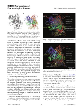
Figure 4. The human PI3K-α protein complex (Protein Data Bank ID.:
6PYS) with its cocrystallized ligand, (3S)-3-benzyl-3-methyl-5-[5-(2-
methylpyrimidin-5-yl)pyrazolo[1,5-a]pyrimidin-3-yl]-1,3-dihydro-2H-
indol-2-one (P5J). (A) Cartoon representation of the raw form; and (B)
minimized-refined form. Red dots in (A) represent water molecules.
Figure 5. A visual representation of potential 6PYS (human PI3K-α
conformations, addressed chain breaks, added missing protein complex) binding site (SiteMap 1).
atoms or residues, assigned bond orders, converted
selenomethionines, and deleted far-water molecules.
In addition, refinement of the cocrystallize ligand
enables the optimization of its positional and thermal
parameters, resulting in a more precise representation
of its interactions within the active site of the receptor
molecule. This optimization accounted for the spatial
arrangement of atoms, bond lengths, bond angles, and
torsional angles of the ligand. Moreover, ligand refinement
allows for the assessment of ligand-receptor interactions,
such as hydrogen bonding, Van der Waals contacts, and
electrostatic interactions. Precise refinement of these
interactions provides insights into the binding affinity,
specificity, and structural basis of ligand recognition.
Furthermore, ligand refinement evaluates the underlying
principles of binding affinity, specificity, and structural
basis of the ligand concerning optimized ligand refinement
parameters, including the assignment of proper bond Figure 6. A visual representation of potential 6PYS (human PI3K-α
protein complex) binding site (SiteMap 2).
orders, generation of accessible tautomers and ionization
states, and prior virtual screening. 50
6PYS protein and P5J ligand is depicted in Figures 5-9.
3.3. Protein-ligand binding pocket identification In each figure, the binding site constituted some ligand
hydrogen bond donor and acceptor regions, along with
The binding pockets/sites of several therapeutic targets are
significantly impacted by protein dynamics. Moreover, sites where hydrophobic interactions could occur. These
51
the structural information of the protein-ligand complex specific properties of the protein significantly influence
can accelerate the optimization process of potential lead ligand binding and interaction.
compounds and help solve problems related to compound The binding site score is a metric used to assess the
selectivity, pharmacokinetics, and patentability. To ensure likelihood or potency of an interaction between a small-
52
specificity and further relay information between active molecule ligand and a protein at a specific binding site.
and allosteric sites, protein-ligand binding is enhanced by It quantifies the propensity of the ligand to bind to the
physical interactions between the binding site residues of local site of the protein, facilitating firm ligand binding
the protein and the ligand. 53 and the formation of a stable complex. The score is
54
Identifying binding sites that predict the concavity usually presented as a numerical value and can be used
where the core scaffold can bind with the protein is essential to rank or prioritize ligands based on their potential
for rational drug design. Binding pocket identification for binding affinity to the protein. It is computed based
Volume 7 Issue 2 (2024) 9 doi: 10.36922/itps.2340