
Page 108 - ITPS-8-1
P. 108
INNOSC Theranostics and
Pharmacological Sciences Steroids in septic cavernous thrombosis
frontal headache, facial pain, fever, neck rigidity, blurred
vision, and photophobia. Her medical history included
chronic obstructive pulmonary disease, hypertension,
and hyperlipidemia. She had no recent travel history or
infection contact.
On examination, she was alert but appeared drowsy. She
had significant exophthalmos in the right eye. Both pupils
were reactive to light, and eye movements were normal. She
also had mild chemosis. Cranial nerves were grossly intact.
She had neck stiffness and a positive Brudzinski sign. Her
muscle power and reflexes were normal. On auscultation,
she had mild wheezing and normal cardiovascular
parameters and had no rashes.
Initial laboratory workup demonstrated a high level
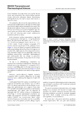
of C-reactive protein (366 mg/L [0 – 10 mg/L]) and white Figure 1. Cranial computed tomography highlighting extensive
blood cell (white blood cells [WBC]; 32.1 × 10^9/L [4 – opacification of the paranasal sinuses – marked opacification of the right
11 × 10^9/L]) and neutrophil [30.8 × 10^9/L (2 – 7.5 × sphenoid (blue arrow), left sphenoid, and ethmoid sinuses consistent with
sinusitis
10^9/L)] counts. Cranial computed tomography (CT)
revealed extensive opacification of the paranasal sinuses
(Figure 1). Given her clinical presentation and laboratory
and imaging findings, the leading differential diagnosis was
sinusitis with a high suspicion of meningeal involvement,
likely bacterial in origin. She was empirically treated with
antibiotics intravenously, particularly chloramphenicol
and cotrimoxazole (she was allergic to penicillin).
Intravenous dexamethasone, 10 mg every 6 h for 4 days,
was also initiated.
On day 2, an ophthalmology consultation was
requested due to persistent exophthalmos, chemosis, and
reduced visual acuity. Subsequent CT venography (CTV)
revealed a small filling defect in the right internal jugular
vein (Figure 2) and right cavernous sinus (Figure 3), which
was suggestive of a thrombus and raised a strong suspicion
of CST. Figure 2. Computed tomography venography. The blue arrow highlights
a small filling defect in the right internal jugular vein that appears darker
Moreover, contrast-enhanced magnetic resonance (hypodense) than the left internal jugular vein, which appears well-
venography (MRV) revealed a filling defect in the right opacified and bright, indicating normal blood flow with contrast filling.
cavernous sinus, confirming the diagnosis of right CST This darker appearance on the right indicates the absence of contrast
(Figure 4). filling in the vein, suggesting thrombus.
Cerebrospinal fluid (CSF) analysis revealed high levels antibiotic therapy with chloramphenicol and cotrimoxazole
of glucose (4.4 mmol/L) and protein (1 g/L), as well as was continued in accordance with institutional protocols
WBC (18 cells/µL) and red blood cell (26 cells/µL) counts, and microbiologist advice.
along with xanthochromia and a negative Gram stain.
CTV and MRV were further reviewed by an interventional The patient’s presentation, laboratory data, and imaging
neuroradiologist, who agreed with the diagnosis of findings were consistent with bacterial meningitis with
meningitis and CST without evidence of subarachnoid secondary CST (SCST). Treatment decisions were made
hemorrhage. A remote neurosurgical consultation was following established protocols for bacterial meningitis
sought, and therapeutic full-dose low-molecular-weight and SCST, which included commencing antibiotics,
heparin (LMWH) was recommended. Blood culture anticoagulation, and dexamethasone.
identified Streptococcus intermedius sensitive to penicillin; By hospital day 4, the patient had shown significant
however, due to the patient’s penicillin allergy, targeted clinical improvement. Her neck pain and headaches
Volume 8 Issue 1 (2025) 102 doi: 10.36922/itps.4853