
Page 11 - JCBP-2-1
P. 11
Journal of Clinical and
Basic Psychosomatics MRI and functional constipation
further discussed WM integrity and structural connectivity have revealed significant increases in ALFF within regions
assessed with DTI. In addition, we illustrated the association associated with the emotional arousal network, such as the
between abnormalities in the brain and constipation anterior insula (aINS), hippocampus (HIPP), dorsal anterior
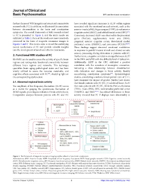
symptoms. The overall framework of MRI research related cingulate cortex (dACC), and orbital frontal cortex (OFC) .
[28]
to FC is presented in Figure 1, and the main results are Conversely, decreased ALFF was observed in the precentral
exhibited in Table 1. Parts of the results are more intuitively gyrus (PreCen), supplementary motor area (SMA),
presented in the form of magnetic resonance images in pregenual anterior cingulate cortex, dorsolateral medial
Figures 2 and 3. This review aims to reveal the underlying prefrontal cortex (DMPFC), and precuneus (PCUN) [28,29] .
neural mechanisms of FC and provide valuable insights These findings suggest abnormal emotional modulation
into the development of novel and effective treatments. in response to painful visceral stimuli and altered somatic
sensory processing during defecation in patients with FC.
2. Functional MRI studies of FC Furthermore, a negative correlation emerged between ALFF
RS-fMRI can be used to assess the activity of specific brain in the SMA and aINS with the difficulty level of defecation.
regions and resting-state functional connectivity between Additionally, ALFF in the OFC exhibited a positive
different brain regions and networks. This technique correlation with the sensation of incomplete evacuation,
quantifies brain aging pathological states and has been indicating a close relationship between dissatisfaction
widely utilized to assess the vascular, metabolic, and with defecation and changes in brain activity, thereby
[27]
cognitive effects associated with FC , shedding light on exacerbating constipation symptoms . Epidemiological
[27]
the corresponding dysfunction. studies, considering a male-to-female patient ratio of 2.1:1,
have examined the impact of gender. Studies have shown
2.1. Abnormal regional brain activity that female patients with FC (FC_F) exhibited lower ALFF
The amplitude of low-frequency fluctuation (ALFF) serves than male patients with FC (FC_M) in the PreCen, thalamus
as a metric for gauging the spontaneous fluctuation of (THA), insula (INS), OFC, ventromedial prefrontal cortex
BOLD signals, providing an indicator of brain activity levels. (VMPFC), and SMA [30,31] . Sex-related differences in brain
Comparative analyses between patients with FC and HC activity revealed that FC_F displays more abnormality in
Figure 1. The framework of magnetic resonance imaging studies related to functional constipation.
Abbreviations: aINS: Anterior insula; ALFF: Amplitude of low-frequency fluctuation; BGN: Basal ganglia network; CCN: Cognitive control network;
dACC: Dorsal anterior cingulate cortex; dFC: Dynamic functional connectivity; DMN: Default mode network; DMPFC: Dorsolateral medial prefrontal
cortex; DTI: Diffusion tensor imaging; FC: Functional constipation; FCAD: Patients with anxiety/depressive disorders; FCDM: Functional connectivity
density mapping; FCNAD: Patients without anxiety/depressive disorders; FNC: Functional network connectivity; GCA: Granger causality analysis;
GM: Gray matter; HC: Healthy control; HIPP: Hippocampus; IFG: Inferior frontal gyrus; LCEN: Left control executive network; MFG: Middle frontal
gyrus; OFC: Orbital frontal cortex; PreCen: Precentral gyrus; RCEN: Right control executive network; RSFC: Resting-state functional connectivity;
SMA: Supplementary motor area; sMRI: Structural magnetic resonance imaging; SN: Salience network; THA: Thalamus; WM: White matter.
Volume 2 Issue 1 (2024) 3 https://doi.org/10.36922/jcbp.1463