
Page 15 - JCTR-10-1
P. 15
Lunevicius et al. | Journal of Clinical and Translational Research 2024; 10(1): 9-17 11
of the liver were other features of the case’s surgical anatomy. Also, hospital. Histopathological investigation of the excised part of
it was the first time an experienced consultant surgeon operated on the gallbladder of 60 × 33 × 24 mm dimensions revealed a 6-mm
a patient with a true left-sided gallbladder. An additional 5-mm wall thickness, necrotic mucosa, and signs of diffuse chronic
diameter port was inserted into the peritoneal cavity through inflammation.
the left upper quadrant of the abdominal wall. An attempt was No other side effects and readmissions to the hospital occurred
made to detach the gallbladder’s fundus from the visceral surface within 90 post-operative days. The patient underwent three-
of segment 3 of the liver. However, this procedure was aborted. dimensional magnetic resonance cholangiopancreatography as an
A decision was made to convert a laparoscopic to open surgery outpatient (Figure 3).
through an upper midline laparotomy. A follow-up visit to the surgical assessment unit on post-
The fundus-first technique was further employed to detach 80% operative day 111 revealed that the patient had made an excellent
of the hepatic wall of the gallbladder from the cystic plate, which post-operative recovery. We used the Gastrointestinal Quality of
was edematous and hemorrhagic. Thereafter, the gallbladder’s Life Index-10 (GIQLI-10, English; point range 0–40; a maximal
fundus was opened, infected bile was suctioned out, and moderate- score indicates perfect health) to assess the quality of life related to
sized gallstones were removed from the cavity of the gallbladder. health [13]. The summative score was 28. However, only diarrhea
When a good backflow of fresh bile was noticed from the internal (score 2 out of 4) had increased since the surgery, which was due
orifice of the cystic duct, situated quite superiorly, a final decision to intake of high-fat or high-sugar foods. The other two low-score
was made to perform a subtotal cholecystectomy. (1 out of 4) symptoms – strong burping/belching and tiredness/
No attempt was made to dissect the cystic pedicle. The
gallbladder was transected circumferentially at the level of the
Hartmann’s pouch. The remnant was closed using two continuous
polyglactin 910 (Vicryl 2/0) and polydioxanone (PDS II 2/0)
®
sutures to obliterate the cavity of the remnant gallbladder.
Floseal , a human gelatine-thrombin matric sealant, was used to
®
ensure hemostasis from the liver. The Portex Robinson drainage
®
system 20 Ch was used for the subhepatic space of the peritoneal
cavity.
2.4. Outcome and follow-up
No surgical complications were observed. However, on post-
operative day 2, a fever episode (38.1°C), supraventricular
tachycardia (>200 beats/min), and hypotension were documented
and managed according to hospital guidelines. Furthermore,
on post-operative day 3, the patient was tested positive for
influenza B. The patient was isolated in a side room with droplet
precautions. The drain was removed from the peritoneal cavity
on post-operative day 6, the day she was discharged from the
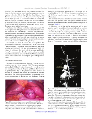
Figure 3. Magnetic resonance cholangiopancreatography (MRCP)
on the 47 post-operative day. The gallbladder remnant is on the right
th
side of the common hepatic and bile ducts, situating adjacent to them.
This image suggests that the fusion of the cystic duct with the common
hepatic duct is on the left of the main bile duct after a U-shaped turn
from right to left anteriorly to the main bile duct. Other anomalies of
the biliary ductal system are highly probable as the right hepatic duct
(RHD) is not identifiable in MRCP images.
Figure 2. Laparoscopic inspection reveals a left-sided gallbladder Abbreviations: ASD: Anterior sectional duct; CBD: Common bile duct;
and acute cholecystitis. The fissure on the visceral surface of the liver CHD: Common hepatic duct; LHD: Left hepatic duct; PD: Pancreatic
between segment 4 of the left hemiliver and segment 5 of the right duct; PSD: Posterior sectional duct; S3: The third segment of the liver;
hemiliver can be interpreted as an external hallmark of the Cantlie- B1: Left-sided duct for caudate lobe; B2, B3, B7, and B8 are segmental
Serege-Rex plane separating the right hemiliver from the left hemiliver. bile ducts; B4, B5, and B6 are not highlighted.
DOI: https://doi.org/10.36922/jctr.00128