
Page 14 - JCTR-10-1
P. 14
10 Lunevicius et al. | Journal of Clinical and Translational Research 2024;10(1) 9-17
2. Materials and Methods 2.2. Differential diagnosis
2.1. Case presentation A working diagnosis of acute calculous cholecystitis was
apparent; however, four diagnostic detail points should be briefly
A right-handed female patient in her 40s with severe overviewed. First, according to Tokyo Guidelines 2007 (TG07),
central epigastric and right-sided hypochondrium pain was 2013 (TG13), and 2018 (TG18), grading the acute cholecystitis
admitted to the emergency general surgery ward of the severity should be emphasized during admission and pre-operative
acute care hospital. The pain was associated with vomiting diagnosis. Our case should have been classified as acute moderate
and diarrhea. Her only concomitant diseases were essential cholecystitis – grade 2 – as it was associated with a duration of
hypertension, which was controlled using 5 mg ramipril daily, acute symptoms of >72 h [10].
and constipation. Her body mass index was 35.8 kg/m on Second, precise radiological characterization of the gallbladder
2
admission. and its site is crucial in managing acute cholecystitis. However,
The patient was afebrile (37.1°C), with a satisfactory oxygen detecting an atypical gallbladder anatomical location is difficult
saturation level at 98%, sinus heart rhythm at 81 beats/min, and (although possible) when performing an urgent transabdominal
high arterial blood pressure at 179/98 mmHg. Further objective ultrasound scan (further details are provided in the discussion).
examination revealed severely tender right hypochondrium, Therefore, the left-sided gallbladder is identified during surgery
central epigastrium, and positive Murphy’s sign. in over 80% of cases [5].
Her white blood cell count was 16.1 × 10 /L. Her neutrophils Third, intraoperative characterization of the anatomy of the
9
comprised 89.4% leukocytes (14.4 × 10 /L). The total serum gallbladder, liver, and its ligaments facilitates decision-making
9
bilirubin concentration was within the standard range (9 µmol/L; during laparoscopic or open surgery. Also, it is essential in
0.53 mg/dL). However, her serum γ-glutamyl transferase education and academic surgery. In the absence of situs viscerum
concentration was 3.8 times above the standard level of inversus, the sinistroposition, a true left-sided gallbladder (our
<40 U/L. Her serum C-reactive protein concentration was within patient), usually with hypoplastic segment 4 of the liver, should
the standard range (4 mg/L). Hyperlactatemia of 3.8 mmol/L be differentiated from the medioposition of the gallbladder [11],
was also detected in the patient. The radiographs did not reveal when it is medially displaced to lie on the undersurface of the
pneumoperitoneum or chest infection. quadrate lobe (i.e., inferior subsegment of segment 4) of the left
The radiologist performed a transabdominal ultrasound hemiliver.
scan within 24 h of admission. Signs of fatty liver disease with Fourth, a right-sided round liver ligament is another rare
hepatomegaly and cholecystolithiasis were reported. Two anatomical variant, which can be associated (but not always; our
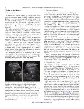
annotated ultrasonograms are illustrated in Figure 1. patient is an example) with the left-sided gallbladder and frequent
intrahepatic vascular and biliary anomalies [12].
A B 2.3. Therapeutic interventions
A standard conservative treatment scheme, including
antibiotics, was established for this patient. We infused 100 mg
of tigecycline and 240 mg of gentamycin through the peripheric
vein, and a regular tigecycline dose of 50 mg every 12 h for
5 days was prescribed. Pyrexia during the hospital stay, local
signs of peritoneal irritation, and serum C-reactive protein
raised to 88 mg/L were key indicators to consider an urgent
index admission laparoscopic cholecystectomy on the 4 day of
th
hospitalization. Informed consent was obtained as a part of the
routine pre-operative actions.
After inserting the first 11-mm diameter port below the
Figure 1. Transabdominal ultrasonography of the gallbladder and umbilicus, a capnoperitoneum of up to 12 mmHg was achieved.
surrounding anatomical structures: (A) longitudinal view of the Standard sites of the right upper quadrant of the abdominal
gallbladder reveals a distended organ and large calculi in its neck; wall were used to insert the other three ports for conventional
the block arrow is directed at the tubular structure which, by our cholecystectomy. Laparoscopic inspection revealed a distended
interpretation, is a branch of the left portal vein; (B) transverse view of thick-walled phlegmonous gallbladder on the left side of the round
the gallbladder shows a calculus within it; most importantly, the head
and the body (the upper block arrow) of the pancreas, and splenic vein and falciform ligaments of the enlarged liver on the anterior wall
behind this organ (the lower block arrow) are visible. A line arrow is of the distal portion of the stomach (Figure 2). A proximal portion
directed at a hypoechogenic area, a site of the round ligament of the of the gallbladder anterior to the hepatic hilum, inflamed tissues of
liver. the hepatoduodenal ligament, unclear segmental anatomy (such as
Abbreviations: LLS: Left lateral section; LPT: Left portal vein; S3: The the presence or absence of segment 4) despite the apparent sulcus
third segment of the liver. on the visceral surface of the liver, and a sizeable umbilical fissure
DOI: https://doi.org/10.36922/jctr.00128