
Page 16 - JCTR-10-1
P. 16
12 Lunevicius et al. | Journal of Clinical and Translational Research 2024;10(1) 9-17
fatigue – were regarded as regular occurrences suffered similarly radiological investigations are restricted to a real-time ultrasound
before subtotal cholecystectomy. scan of the gallbladder [17]. Table 1 describes why it is difficult
to reveal a left-sided gallbladder through standard examination
3. Discussion and imaging techniques before surgery [3,18-22]. On the
The primarily aims of surgical care are to save the patient’s contrary, a left-sided gallbladder and the variations of the biliary
life, prevent the patient from further disease complications or tract – a frequent combination of biliary anomalies – can be
reduce the risk of sustaining them, improve the patient’s quality diagnosed preoperatively using intravenous contrast-enhanced
of life, and eliminate the possibility of iatrogenic injury associated reconstructive three-dimensional computed tomography
with surgery. Gallbladder surgery for benign biliary disease is an (CT)-cholangiography [23-25]. However, a three-dimensional
CT-cholangiography is not a routine investigation in an acute
excellent example of this concept because injury to any classified care surgery environment. It can be considered when a congenital
bile duct is considered avoidable [14-16]. This paper highlights anomaly of the gallbladder is suspected during an ultrasound scan
the decision-making during and the technical details of gallbladder examination. The same logic is relevant for applying an urgent
surgery related to double conversion in an acute surgery setting magnetic resonance cholangiopancreatography.
with atypical gallbladder anatomy. Conversions from laparoscopic Second, a targeted laparoscopic inspection of the liver and
to open surgery and pre-planned total to subtotal cholecystectomy gallbladder through a first port and the rationale for correctly
with the closure of the gallbladder remnant guaranteed no using other laparoscopic ports and instruments are fundamental
intraoperative risks, satisfactory surgical outcomes, and effective principles of safe laparoscopic surgery for all, as an element
physical rehabilitation following the arduous gallbladder surgery. of uncertainty is a satellite of every surgery. Unfortunately, the
Seven other themes related to the left-sided gallbladder –precision in gallbladder anatomy-related intraoperative problem was not
radiological diagnostics, the importance of laparoscopic inspection, identified and acknowledged during the primary inspection of the
detailed informed consenting, extraordinarily high bile duct injury hepatobiliary area. This determined the standard insertion of the
rates, variations of ductal anatomy, intraoperative fluorescent other three laparoscopic ports through the right upper quadrant of
cholangiography, and decision-making to perform a less-than-total the abdominal wall. If the problem had been identified during the
gallbladder removal – emerged from the details of this case report. primary inspection, the second port would have been inserted into
First, pre-operative identification of the left-sided gallbladder the peritoneal cavity through the left lateral quadrant laterally to
is difficult, especially in emergency admission patients whose create an adequate workspace between the round and falciform
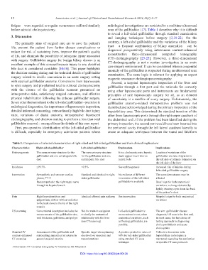
Table 1. Comparison of selected characteristics of right-sided and left-sided gallbladders and their clinical implications
Characteristics Right-sided gallbladder Left-sided gallbladder Explanation Implications
Embryogenesis The same primary structure for the The same primary structure It is a cholecystic axis; hepatic Locational variations of the
gallbladder and one extrahepatic bile for the gallbladder and one ducts appear much later as gallbladder are rare: migration to
duct extrahepatic bile duct lateral buds the left side or primary formation on
the left side of the liver
Incidence ≥99.7% <0.3% See embryogenesis Increased risk of injuries during
left-sided gallbladder surgery
Innervation Sympathetic and sensory: coeliac Standard and identical to right- No evidence of different The same dermatomes may be
plexus, T7–9 sided gallbladder innervation of the left-sided affected
Parasympathetic: the right vagus nerve gallbladder is available Boas’ sign for both anatomical
through its hepatic branch variations: a change detected by
lightly drawing a pin down the back
of the patient’s chest
Pain Right hypochondrium and Identical afferent pain pathway See innervation Murphy’s sign for both anatomical
epigastrium, with or without radiation variations
to the back close to the tip of the right
scapula
US scanning Conventional description includes the Not the main investigation Left-sided gallbladder is The aim: gallbladder disease
measurements of the gallbladder size, to clarify the anatomical an occasional event; other diagnosis; US scan is the first and,
wall thickness, gallstones, and polyps relationship with the liver anatomical variations, such in most cases, the last choice of
(see CT scanning) as floating gallbladder, are testing approach to diagnosing
possible. cholecystolithiasis and acute
cholecystitis
Standard IV Assessment of the gallbladder and Specific target when planning A positive predictive value of Collective discussion with
contrast-enhanced surrounding anatomical structures in elective liver resection and 60% for left-sided gallbladder hepatobiliary radiologists is
CT scanning general surgical practice transplantations using standard CT scan warranted regarding the application
technique of specific CT scan protocols
Abbreviations: CT: Computed tomography; IV: Intravenous; US: Ultrasound
DOI: https://doi.org/10.36922/jctr.00128