
Page 17 - JCTR-10-1
P. 17
Lunevicius et al. | Journal of Clinical and Translational Research 2024; 10(1): 9-17 13
ligaments, left lateral section of the liver, and the gallbladder [25]. from injuries to the highly probable anomalous extrahepatic bile
The location of the third, fourth, and (if the need arises) fifth ducts.
ports for traction of the gallbladder should be adapted according Variations of biliary anatomy at the hepatic hilum are more
to the anatomical situation and surgeons’ preferences. This point frequent in patients with left-sided gallbladder, especially in
should be regarded as a reminder to the surgeons to inspect the those with abnormal intrahepatic portal vein branching [23].
liver and gallbladder after the insertion of the laparoscope through The understanding of infraportal bile duct anatomy, classified
the periumbilical port and early recognize the abnormal position as joining the hepatic duct caudally to the transverse portion
of the gallbladder to allow the standard port placements to be of the left portal vein [30], is of paramount importance for
modified [26]. safe cholecystectomy planning. A few variations in infraportal
Third, the theoretical reasons for performing alternative courses of segmental and sectional bile ducts were reported.
gallbladder surgeries should be discussed with the patient They should be considered before, as it is possible to identify
comprehensively for informed consent [27]. The options for them using contrast-enhanced computed tomography and
managing a left-sided gallbladder were not discussed with our magnetic resonance-based imaging, and during gallbladder
patient preoperatively. Interestingly, the incidences of a left-sided surgery. The examples include infraportal B1l (it is one
gallbladder (not routinely discussed while providing information of the bile ducts of segment 1 which drains Spiegel’s lobe)
for informed consent) and major bile duct injury (discussed joining the left or common hepatic duct [30], right posterior
routinely) are similar. It is approximately 0.3%. sectional bile duct joining the right anterior sectional bile duct
Fourth, comparisons of bile duct injury rates from both reviews with an infraportal course [31], right posterior sectional duct
on cholecystectomy for a left-sided gallbladder [5,6] with the joining the common bile duct [32], and infraportal B3 [33].
CholeS Study Group [28] data for conventional cholecystectomy, Encountering another infraportal bile duct of the left hemiliver
are concerning. For example, four patients in the cholecystectomy is always possible, as a true left-sided gallbladder is more
for a left-sided gallbladder cohort had an injury to the bile duct associated with the left-sided biliary tract variations. Thus,
with a rate of 7.3% [6], which is 4.3 times higher than the bile infraportal variations of biliary anatomy at the hepatic hilum
duct injury rate (1.7%) for the most difficult grade 4 and 5 are the second reason a surgeon should initiate the dissection
cholecystectomies. Furthermore, it is almost 43 times higher than of the left-sided gallbladder as close to its wall as possible to
the bile duct injury rate (0.17%) for grade-3 difficulty-specific prevent infraportal bile duct injury [24]. It is a prerequisite for
cholecystectomies and 29 times higher than the overall bile duct safe total cholecystectomy.
injury rate of 0.25% in the CholeS Study [28]. Such comparisons Sixth, an intraoperative fluorescent cholangiography method
have methodological drawbacks; nonetheless, they indicate that a using indocyanine green and a near-infrared light source is a new
left-sided gallbladder and associated variations in biliary ductal imaging method in laparoscopic cholecystectomy to improve the
anatomy present challenges in intraoperative decision-making and visualization of the extrahepatic biliary anatomy (despite a long
the technical execution of the surgical procedure [29]. history of indocyanine green utilization in liver surgery) [34].
Fifth, the atypical position of the gallbladder predetermines the However, it should be noted that surgical care providers can
cystic duct’s atypical anatomical relationship with the main bile use intraoperative imaging methods approved by the individual
ducts, first- and second-order bile ducts, and the entire hepatic health-care organization.
pedicle. Specifically, the left-sided gallbladder, anterior to the Seventh, when in doubt, an anatomic dissection of the
hepatic pedicle, changes Calot’s triangle planes from horizontal proximal portion of the gallbladder and cystic pedicle cannot be
and lateral to vertical and anterior, bringing the gallbladder closer performed safely, or the hepatic wall of the gallbladder cannot
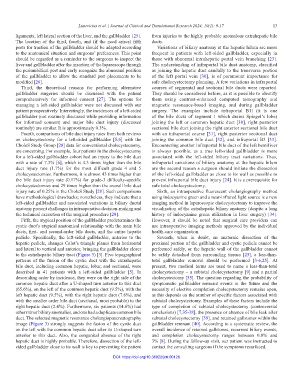
to the extrahepatic biliary tract (Figure 3) [3]. Five topographical be safely detached from surrounding tissues [25], a less-than-
patterns of the fusion of the cystic duct with the extrahepatic total gallbladder removal should be performed [3-6,25]. At
bile duct, including common hepatic, lobar, and sectional, were present, two medical terms are used to name a less-than-total
described in 41 patients with a left-sided gallbladder [5]. In cholecystectomy – a subtotal cholecystectomy [9] and a partial
descending order by incidence, they were on the right side of the cholecystectomy [35]. The question regarding the probability of
common hepatic duct after a U-shaped turn anterior to this duct symptomatic gallbladder remnant events in the future and the
(65.6%), on the left of the common hepatic duct (9.5%), with the necessity of elective completion cholecystectomy remains open,
left hepatic duct (9.5%), with the right hepatic duct (7.6%), and as this depends on the number of specific factors associated with
with the smaller order bile duct (sectional, most probable) to the subtotal cholecystectomy. Examples of these factors include the
right hepatic duct (2.4%). Furthermore, six patients (14.6%) had type of completion of subtotal cholecystectomy (controversial
other minor biliary anomalies, and one had a duplicate common bile conclusions) [7,35-38], the presence or absence of bile leak after
duct. The selected magnetic resonance cholangiopancreatography subtotal cholecystectomy [39], and retained gallstones within the
image (Figure 3) strongly suggests the fusion of the cystic duct gallbladder remnant [40]. According to a systematic review, the
on the left with the common hepatic duct after its U-shaped turn overall incidence of retained gallstones, recurrent biliary events,
anterior to this duct. Also, the congenital absence of the right and completion cholecystectomy ranges between 0.8% and
hepatic duct is highly probable. Therefore, dissection of the left- 3% [8]. During the follow-up visit, our patient was instructed to
sided gallbladder close to its wall is key to preventing the patient contact the consulting surgeons if the symptoms resurfaced.
DOI: https://doi.org/10.36922/jctr.00128