
Page 32 - JCTR-10-1
P. 32
28 Shah et al. | Journal of Clinical and Translational Research 2024; 10(1): 25-32
7.3 mg/dL (direct – 5.8 mL/dL), ALT – 88 IU/L, AST – 65 IU/L,
ALP – 104 IU/L, and GGTP – 92 IU/L. Prednisolone was tapered
over the next 10 weeks and stopped after complete normalization
of liver function. He was further followed for 10 weeks after
steroid discontinuation, during which recurrence did not occur.
3.2.3. Case 3 - Dapsone-induced liver injury
A 52-year-old male patient presented with worsening jaundice
and pruritus for the 1.5 months before seeking medical consultation
at our hospital. He had been taking dapsone 100 mg daily as anti-
leprosy treatment in the past 3 months. He was not taking any other
medications, was a non-addict, and had no comorbidities, except
leprosy. On physical examination, madarosis, contracture of upper
limb fingers, and large hypopigmented hypoesthetic patches at the
trunk and back were present. Features of hypersensitivity were
absent. He had deep icterus but there were no clinical signs of liver
failure. TB and DB were 49 and 40 mg/dL, respectively. KF rings
were present in both eyes, and 24-h urinary copper was slightly
elevated (Table 3). Serum ceruloplasmin was normal. His RUCAM
score was 6 points, and his R-value was 2.4, suggesting a mixed
pattern of DILI. Based on these results, dapsone was discontinued,
and a liver biopsy was performed, demonstrating prominent acinar
disarray, mild-to-moderate inflammatory infiltrates in the portal
tract, and ductular reactions with focal neutrophilic cholangitis.
Giant hepatocytes, zone 3 canalicular and intrahepatic cholestasis,
and prominent zone 3 perivenulitis were also noted. Copper staining
was negative. The overall picture suggested mixed hepatitis and
cholestatic pattern, which was possibly drug-induced (Figure 3,
Case 3). Emollients, anti-histaminic, and UDCA were given
but liver functions continued to worsen, and oral prednisolone
(40 mg/day) was started as rescue therapy. At 2 weeks of follow-
up, his TB was 8.3 mg/dL (direct – 6.6 mL/dL). Other liver function
parameters were ALT – 65 IU/L, AST – 98 IU/L, ALP – 154 IU/L,
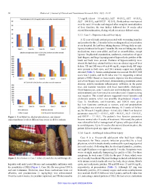
Figure 1. Total bilirubin, alkaline phosphatase, and alanine and GGTP – 171 IU/L. The patient’s liver function parameters
aminotransferase levels at different time points in all five patients. became normal after 8 weeks of treatment. Afterward, the patient
was referred for further management of leprosy with special advice
A B to avoid dapsone. Up to 18 weeks after steroid discontinuation, the
patient did not report any signs of recurrence.
3.2.4. Case 4 - Antifungal-induced liver injury
Case 4 is a 16-year-old adolescent who had been taking
itraconazole for Tinea corporis infection prescribed by a local
physician, which he inadvertently continued for a prolonged period
(several weeks). Following this, he developed jaundice, pruritus,
and night blindness over approximately 3 weeks. Symptoms were
worse at night, markedly hampering his quality of life. For these
symptoms, he took some CAMs for the past 10 days, which were
Figure 2. Skin lesions in Case 1 before (A) and after steroid therapy (B). not clinically beneficial. Physical findings included exfoliated skin
with intense scratch marks all over the body, deep icterus, Bitot’s
hepatitis with mild portal fibrosis and eosinophilic infiltrates with spots, and ecchymotic patches. TB and DB were 15 and 10 mg/dL,
the possibility of DILI (Figure 3, Case 2b). He was given UDCA respectively, at presentation (Table 3). Although KF ring was
and anti-histaminic. However, since these medications were not bilaterally positive, 24-h urinary copper and serum ceruloplasmin
effective, oral prednisolone (1 mg/kg/day) was administered. were normal. His RUCAM score was 6 points, and his R-value was
Over the next 4 weeks, his pruritus improved, and TB decreased to 2.4, indicating a mixed pattern of DILI. He had severe cholestasis
DOI: https://doi.org/10.36922/jctr.00104