
Page 98 - JCTR-10-1
P. 98
94 Mardany et al. | Journal of Clinical and Translational Research 2024; 10(1): 93-98
Some studies supported the association between skeletal pattern the sella turcica), N (the intersection points of the nasion and the
and the airway, while others did not show such a relationship. frontal bone in the sagittal view), A (the innermost point on the
A study by Jadhav et al. [6] demonstrated that there was no anterior contour of the maxilla below the maxillary plane), and B
significant correlation between the total airway volume and three (the innermost point on the anterior mandibular shape above the
sagittal skeletal groups. Alhammadi et al. [7] reported that the pogonion). The Wits appraisal is the measured distance between A
volume of the palatopharyngeal and glossopharyngeal airways and B along the mid-sagittal reference line. GoGn-SN angle was
and the narrowest point of the palatopharyngeal airway were measured between the line of the gonion (Go) and gnathion (Gn)
greater in Class II skeletal than in other skeletal groups. Alves and the sella-nasion (SN) line. All patients were middle easterners
et al. [8] found that the type of malocclusion did not influence and had normal growth patterns.
the dimensions and volumes of the airway in most cases. On All CBCT images were selected from patients with a mandibular
the other hand, Tseng et al. [9] showed that individuals with plane angle of 32 ± 5 = GoGn-SN.
Class II skeletal malocclusion have smaller airway volumes ● Class I: 0° < ANB < 4°; −1 mm < Wits < 0 mm
than individuals with Class I and III malocclusion. In a study ● Class II: ANB > 4°; 0 mm < Wits
by Shokri et al. [10], it was shown that the volume and area of ● Class III: ANB < 0°; Wits < −1 mm
the airway were significantly greater in Class III patients than Exclusion criteria of this study are as follows:
in Class I or II. Zeng et al. [11] demonstrated that the volume The patients who had no history of orthognathic surgery,
of the pharyngeal airway was significantly greater in Class III nasal surgery, syndromes, trauma, or pathology in the airway and
and Class I patients compared to Class II patients. Due to pharynx.
the significant discrepancies in the results among the studies CBCT images that lacked diagnostic value.
mentioned above and the lack of research regarding different CBCT images were converted to DICOM format and transferred
sagittal malocclusion with normal growth patterns in the Middle to 3D Dolphin software (Management & Imaging Solutions,
Eastern population, the necessity of conducting this research Chatsworth, CA, USA). The overall volume of the pharyngeal
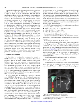
became evident. Therefore, this study aimed to measure the airway and the most constricted area (mm ) were assessed and
2
relationship between the airway volume and skeletal Class I, determined (Figures 1 and 2). The measurements were performed
Class II, and Class III malocclusions with normal growth by two researchers, and the intraclass correlation coefficient (ICC)
patterns in individuals aged 17 – 39 years using CBCT. was calculated to determine the reliability of the two researchers.
2. Patients and Methods In this study, the ICC was above 80%, indicating the reliability of
the two researchers.
In this study, we conducted a comprehensive analysis of The definitions used throughout this study are as follows:
CBCT obtained from the Department of Oral and Maxillofacial
Radiology archives at Tehran Medical Sciences, Islamic Azad 2.1. Total pharyngeal airway volume (TP)
University. The survey was conducted in accordance with the The upper bound of the pharyngeal airway passes through PNS
guidelines of the Declaration of Helsinki. All human research and is parallel to the standard horizontal plane; the lower bound
was conducted in accordance with the ethical standards of the passes through C4 and is parallel to the standard horizontal plane.
committee responsible for human experimentation (institutional
and national), and with the Helsinki Declaration of 1975, revised
in 2013. Ethical approval was obtained from the Islamic Azad
University Local Research Ethics Committees (protocol identifier
IR.IAU.DENTAL, REC; 1400.041).
In this cross-sectional analytical study, the 90 CBCT images
were divided into three groups, with 30 patients in each class,
namely Class I, II, and III malocclusions. These CBCT images
were obtained using a Sirona Galileos Sirona Dentsply device in
Germany; all images were prepared by the Scan-Fast protocol, with
a scan time of 14 s, a field of view of 15 cm × 15 cm, 98 kV, and
3 mA. All CBCTs were performed when the patients assumed the
standing position, and patients stood when looking at themselves
in the mirror. All images were taken from CBCT scans where the
teeth were in occlusion, and all cephalograms of CBCT scans are
completely real because they were extracted from CBCT images
captured using the Sirona Galileos device, Germany. The patients Figure 1. Three-dimensional upper airway model.
were divided into three groups: Class I, Class II, and Class III, Abbreviations: VP: Velopharyngeal airway volume;
based on the ANB angle and Wits appraisal. The SNA and SNB GP: Glossopharyngeal airway volume; OP: Oropharyngeal volume;
angles were measured using the following points: S (the center of TP: Total pharyngeal airway volume.
DOI: https://doi.org/10.36922/jctr.23.00110