
Page 18 - JCTR-10-6
P. 18
328 Barreto et al. | Journal of Clinical and Translational Research 2024; 10(6): 325-333
A
B
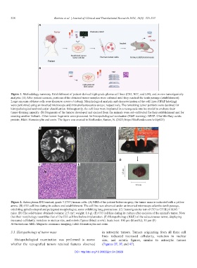
Figure 1. Methodology summary. Establishment of patient-derived high-grade glioma cell lines (C03, N07, and L09), and in vivo tumorigenicity
analysis. (A) After patient consent, portions of the obtained tumor samples were cultured until they reached the tenth passage (establishment).
Large amounts of these cells were frozen to create a biobank. Morphological analysis and characterization of the cell lines (GFAP labeling)
were performed using an inverted microscope and immunofluorescence assays, respectively. The remaining tumor portions were destined for
histopathological and molecular classification. Subsequently, the cell lines were implanted in a xenogeneic murine model to evaluate their
tumor-forming capacity. (B) Fragments of the tumors developed and excised from the animals were sub-cultivated for their establishment and for
creating another biobank. Other tumor fragments were processed for histopathological evaluation (H&E staining). GFAP: Glial fibrillary acidic
protein; H&E: Hematoxylin and eosin. The figure was created in BioRender; Santos, N. (2023; https://BioRender.com/w44p823)
A B C
D
F E
Figure 2. Astrocytoma IDH-mutant, grade 3 (C03) human cells. (A) MRI of the patient before surgery; the tumor mass is indicated with a yellow
arrow. (B) C03 cell line during its culture and establishment. The cell line was observed under an inverted microscope after the tenth passage,
exhibiting spindle-shaped and polygonal morphologies; some exhibiting long protrusions. (C) Tumorigenicity test of C03 in C57BL/6 RAG
−/−
3
mice. (D) The solid tumor obtained (volume: 2.5 cm ; weight: 1.6 g). (E) C03 cell line during its culture after excision of the animal’s tumor. Note
that their morphology resembles that of the C03 cell line before implantation. (F) Histopathology (H&E) of the subcutaneous tumor, displaying
increased cellularity, variation in nuclear size, and mitotic figures (black arrow). Scale bars: 100 µm (B and E); 50 µm (F)
Abbreviations: MRI: Magnetic resonance imaging; H&E: Hematoxylin and eosin
3.3. Histopathology of tumor mass in astrocytic tumors. Tumors originating from all three cell
lines indicated increased cellularity, variation in nuclear
Histopathological examination was performed to assess size, and mitotic figures, similar to astrocytic tumors
whether the xenografted tumors retained features observed (Figures 2F, 3F, and 4F).
DOI: http://doi.org/10.36922/jctr.24.00028