
Page 20 - JCTR-10-6
P. 20
330 Barreto et al. | Journal of Clinical and Translational Research 2024; 10(6): 325-333
A B C D
E F G H
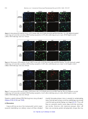
Figure 5. Astrocytoma IDH-mutant, Grade 3 (C03) human cells. (A-D) Phalloidin probe and GFAP labeling. The cells displayed rounded
morphology with some thin extensions; GFAP staining was positive in C03 cells, confirming astrocytic origin. (E-H) Negative control
(without GFAP labeling). Scale bars: 100 µm
A B C D
E F G H
Figure 6. Glioblastoma IDH-wildtype, Grade 3 (N07) human cells. (A-D) Phalloidin probe and GFAP labeling. The cells displayed rounded
morphology with some thin extensions; GFAP staining was positive in N07 cells, confirming astrocytic origin. (A-H) Negative control
(without GFAP labeling). Scale bars: 100 µm
A B C D
E F G H
Figure 7. Astrocytoma IDH-mutant, grade 3 (L09) human cells. (A-D) Phalloidin probe and GFAP labeling. The cells displayed rounded
morphology with some thin extensions; GFAP staining was positive in L09 cells, confirming astrocytic origin. (E-H) Negative control
(without GFAP labeling). Scale bars: 100 µm
Negative controls (without GFAP labeling) were also performed directly from patient tumor tissue is essential to understanding
(Figures 5E-H, 6E-H, and 7E-H). tumor behavior and biology, as well as establishing a preclinical
4. Discussion model for testing and developing new drugs [10,18]. These cell
lines are commonly used to assess drug sensitivity, resistance,
Cancer cell lines are one of the most powerful tools in cancer and toxicity. However, after numerous passages, these cell
research. Establishing new primary cancer cell lines obtained lines can accumulate genetic and epigenetic changes that may
DOI: http://doi.org/10.36922/jctr.24.00028