
Page 62 - JCTR-11-2
P. 62
Journal of Clinical and
Translational Research CT-guided needle versus glue localization
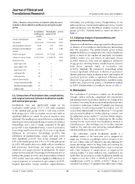
Table 1. Baseline characteristics of patients and pulmonary lobectomy) and pathology results (benign lesions, in situ
nodules in the localization needle group and medical glue adenocarcinoma, microinvasive adenocarcinoma, invasive
group adenocarcinoma) were distributed similarly between the
groups (p>0.05). Detailed statistical results are shown in
Localization Medical glue p‑value
needle group group (n=22) Table 2.
(n=64)
Gender (male/female) 18/46 6/16 0.939 3.3. Subgroup analysis of pneumothorax and
pulmonary hemorrhage
Age (years) 52.45±12.43 52.35±10.55 0.922
Smoking history (yes/no) 10/54 3/19 0.822 Patients were divided into subgroups based on the presence
or absence of pneumothorax and pulmonary hemorrhage
Pulmonary disease history 10/54 4/18 0.748 after the procedure. The pneumothorax group showed
(yes/no) significant differences compared to the nonpneumothorax
Nodule size (mm) 12.40±4.33 13.77±5.41 0.235 group in terms of the number of punctures, localization
Distance to pleura (mm) 13.247±10.01 11.159±7.89 0.381 method, nodule size, and history of pulmonary disease
Nodule location 0.661 (p<0.05). However, there were no significant differences
Right upper lobe 24 9 in age, gender, smoking history, nodule location, distance
Right middle lobe 3 1 from pleura, puncture depth, or localization time
Right lower lobe 9 5 (p>0.05). Similarly, the pulmonary hemorrhage group
Left upper lobe 18 3 revealed significant differences in history of pulmonary
disease, puncture depth, localization time, and number of
Left lower lobe 10 4 punctures (p<0.05), while no significant differences were
Nodule characteristics 0.725 observed in age, gender, smoking history, nodule location,
Pure ground-glass nodules 22 5 nodule size, distance from pleura, or localization method
Mixed ground-glass nodules 34 12 (p>0.05). Detailed statistical results are shown in Table 3.
Solid nodules 8 5
4. Discussion
3.2. Comparison of localization time, complications, The localization of pulmonary nodules can be achieved
and surgical outcomes between localization needle through various methods, categorized into preoperative
and intraoperative localization based on the timing of the
and medical glue groups
procedure. Currently, the most commonly used preoperative
Localization time was significantly longer in the localization techniques include CT-guided percutaneous
localization needle group (17.19 ± 2.56 min) compared localization, bronchoscopy-assisted localization, and CT
to the medical glue group (15.36 ± 2.06 min, p<0.05). virtual three-dimensional (3D) localization. 14,15 CT-guided
Both groups had a 100% localization success rate, with no percutaneous localization involves placing a marker near
significant differences noted. No pleural reactions were the pulmonary nodule under CT guidance before surgery.
observed. The needle group reported more complications, The nodule is then located by searching for the marker,
including 15 cases of pneumothorax, 11 cases of pulmonary making this the most widely used localization method in
hemorrhage, and 1 case of cough, compared to the glue clinical practice. This technique can be further classified
group with 1 case of pneumothorax, 2 cases of pulmonary based on the materials used, including medical adhesive,
hemorrhage, and 5 cases of cough. There was no significant lipiodol, autologous blood, and others. 16-18
difference in pulmonary hemorrhage (p>0.05), but The pulmonary nodule localization needle is a
differences in pneumothorax, cough, and pain scores modified version of the hook-wire and is designed for
were significant (p<0.05). Pain scores averaged 2.89 ± single use. It features a quadruple-barbed structure at the
0.95 in the needle group and 2.36 ± 0.79 in the glue group tip and a three-color soft thread at the tail, allowing for
(p<0.05). All complications were mild, with pneumothorax secure anchorage within the lung tissue and marking the
and pulmonary hemorrhage not requiring emergency depth of localization. This method has a high success rate
treatment, and coughs relieved by antitussives. No severe and a low incidence of complications, as supported by
complications occurred. The average VATS time was similar existing studies. A study indicates that, compared to the
19
between the groups (p>0.05), and all procedures achieved hook-wire, the pulmonary nodule localization needle has
100% nodule resection, with no significant differences in a higher success rate (100% vs. 84.0%, p<0.05) and fewer
20
surgical approach or conversion to open surgery (p>0.05). complications (12.9% vs. 40.0%, p<0.05). Wang et al.
Surgical approaches (wedge resection, segmentectomy, reported that using a CT-guided novel pulmonary nodule
Volume 11 Issue 2 (2025) 56 doi: 10.36922/JCTR025070007