
Page 63 - JCTR-11-2
P. 63
Journal of Clinical and
Translational Research CT-guided needle versus glue localization
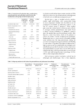
Table 2. Comparison of localization time, complications, localization needle before thoracoscopic resection of SPNs
surgical approach, and pathology results between the effectively reduces the risk of displacement or dislodgement
localization needle group and medical glue group of the hook-wire and alleviates postoperative pain. 21
Localization Medical p‑value Medical glue, a type of medical adhesive product,
needle group glue group enables rapid solidification, with cyanoacrylate being
(n=64) (n=22) its main component. Scholars first described its use for
Localization time (min) 17.19±2.56 15.36±2.06 0.003 preoperative localization of pulmonary nodules in the
Number of punctures (1/>1) 44/20 4/18 0.238 late 20 century, using CT guidance. When medical
th
22
Complications glue comes into contact anionic substances such as blood,
Pneumothorax (yes/no) 15/49 1/21 0.049 it undergoes anionic polymerization and rapidly forms
23
Pulmonary hemorrhage 11/53 2/20 0.501 a clump. During localization, CT guidance is used to
(yes/no) inject the medical glue near the pulmonary nodule. After
Cough (yes/no) 1/63 5/17 0.004 injection, a hard mass forms, which can be palpated to
Pain score 2.89±0.95 2.36±0.79 0.021 confirm its position, thereby completing the localization.
Vats time (min) 114.6±35.1 116.6±34.2 0.819 Medical glue acts as a tissue sealant that rapidly solidifies
Surgical approach 0.086 upon injection into the lung parenchyma, sealing the
puncture tract and preventing pneumothorax and bleeding
Wedge resection 41 14 to some extent. CT-guided medical glue localization has a
Segmentectomy 16 2 high success rate and low complication rate, making it a safe
Lobectomy 7 6 and effective preoperative localization method. Studies have
Pathology results 0.315 reported localization success rates of up to 100% and low
Benign lesions 11 2 complication rates, including asymptomatic pneumothorax
In situ adenocarcinoma 5 4 (11.9%) and mild intrapulmonary hemorrhage (13.1%),
Microinvasive 11 6 with no other complications observed. 24
adenocarcinoma Each pulmonary nodule localization method has its
Invasive adenocarcinoma 37 10 advantages and disadvantages. Due to their simplicity,
Table 3. Subgroup analysis of risk factors for pneumothorax and pulmonary hemorrhage
Pneumothorax Non‑pneumothorax p‑value Pulmonary Non‑pulmonary p‑value
group (n=16) group (n=70) hemorrhage group hemorrhage group
(n=13) (n=73)
Age (years) 53.73±13.61 52.18±11.61 0.65 54.15±8.79 52.15±12.41 0.58
Gender (male/female) 3/13 21/49 0.539 6/7 18/55 0.111
Smoking history (yes/no) 1/15 12/58 0.446 4/9 9/64 0.103
Pulmonary disease history (yes/no) 6/10 8/62 0.011 5/8 9/64 0.019
Nodule location 0.105 0.434
Right upper lobe 2 31 5 28
Right middle lobe 2 2 0 4
Right lower lobe 4 10 4 10
Left upper lobe 5 16 3 18
Left lower lobe 3 11 1 13
Nodule size (mm) 10.63±2.12 13.24±4.91 0.002 13.15±5.09 12.68±4.58 0.739
Distance to pleura (mm) 14.03±10.93 12.44±9.32 0.561 15.80±11.73 12.163±9.12 0.209
Puncture depth (mm) 18.89±10.54 18.22±7.58 0.773 23.07±9.41 17.49±7.61 0.021
Localization time (min) 17.00±2.20 16.66±2.63 0.644 18.46±1.85 16.41±2.55 0.007
Localization method (needle/glue) 1/15 49/21 0.049 11/2 53/20 0.499
Number of punctures (1/>1) 6/10 56/14 0.001 6/7 56/17 0.04
Volume 11 Issue 2 (2025) 57 doi: 10.36922/JCTR025070007