
Page 41 - JCTR-9-4
P. 41
Sondore et al. | Journal of Clinical and Translational Research 2023; 9(4): 253-260 257
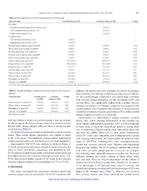
Table 4. Procedural and VH-IVUS characteristics of PCI group
Characteristic Carotid artery (n=22) Coronary artery (n=22) P‑value
PCI artery
Left anterior descending coronary artery, n (%) - 8 (36.4) -
Left circumflex coronary artery, n (%) - 4 (18.2) -
Right coronary artery, n (%) - 10 (45.5) -
Analyzed artery
Left internal carotid artery, n (%) 6 (27.3) - -
Right internal carotid artery, n (%) 16 (72.7) - -
Minimal lumen diameter, mm, mean±SD 3.6±0.6 2.0±0.7 <0.01
Mean lumen diameter, mm, mean±SD 5.0±0.5 2.9±0.1 <0.01
Minimal lumen area, mm, mean±SD 13.8±3.9 4.2±0.3 <0.01
Minimal vessel diameter, mm, mean±SD 6.3±1.0 3.8±0.1 <0.01
Mean vessel diameter, mm, mean±SD 7.4±0.8 4.5±0.2 <0.01
Vessel volume, mm , mean±SD 716.7±447.9 309.6±37.7 <0.01
3
Plaque volume, mm , mean±SD 390.3±268.5 182.3±24.0 <0.01
3
Plaque burden, %, mean±SD 53.4±7.7 57.8±5.8 0.03
Segment length, mm, mean±SD 15.4±8.3 18.1±1.5 0.17
Necrotic tissue, %, mean±SD 18.6±9.4 22.5±1.9 0.05
Fibrotic tissue, %, mean±SD 56.5±8.0 50.6±2.1 0.07
Fibrolipids, %, mean±SD 19.3±10.2 13.9±2.7 0.36
Calcium, %, mean±SD 5.6±4.4 13.6±1.8 <0.01
VH-IVUS: Virtual histology intravascular ultrasound, PCI: Percutaneous coronary intervention, SD: Standard deviation
Table 5. Virtual histology comparison between carotid and coronary multiple risk factors who were scheduled for carotid or coronary
arteries artery stenting. The findings of the present study were as follows:
Characteristic Carotid artery Coronary P‑value (1) the carotid plaque composition was significantly correlated
(n=100) artery (n=100) with coronary plaque phenotype; (2) the percentage of NC and
Necrotic tissue, %, mean±SD 19.8±9.4 22.6±7.3 <0.01 calcium tissue was significantly higher in the coronary arteries,
Fibrotic tissue, %, mean±SD 53.4±8.0 51.7±10.3 0.09 whereas the amount of FF plaque component was greater in the
Fibrolipids, %, mean±SD 19.6±9.6 12.5±9.1 <0.01 carotid arteries; and (3) patients with a history of cerebrovascular
Calcium, %, mean±SD 7.7±5.6 13.6±8.2 <0.01 events had a tendency of greater proportion of NCs in their carotid
SD: Standard deviation arteries compared to others in CAS group.
Inflammation in atherosclerotic plaque produces systemic
with and without a history of cerebrovascular event. In contrast, effects, thus, active, ongoing inflammation at one vascular bed
the percentage of all analyzed tissue types in the coronary arteries could enhance inflammation in another. A NC in VH corresponds
did not differ among patients with and without cerebrovascular to tissue areas in which the extracellular matrix is lacking (total
event histories (Table 6). loss of supporting collagen) and has been replaced by dead cells
Two patients had post-procedural acute ipsilateral cerebrovascular and lipid-rich cellular debris [10,11], thus, tissue characterized
events. Their carotid plaque composition was similar to others as the NC is the most vulnerable part of atherosclerotic lesions
in the CAS group. Non-ipsilateral post-procedural intrahospital with ongoing inflammation. We observed that all analyzed tissue
cerebrovascular events were not observed in our study population. types by VH-IVUS, including NCs, were correlated between
Representative VH-IVUS case analysis is shown in Figure 2, carotid and coronary vascular beds. Together with knowledge
in which increased necrotic tissue amount in carotid and coronary from previous studies, VH-IVUS analysis confirms that patients
artery of 74.0% and 48.8%, respectively, was identified by VH- with more vulnerable plaque patterns at one site are at higher
IVUS analysis of culprit lesion in the left internal carotid artery and risk of having more vulnerable plaques at another site. However,
non-culprit lesion in the mid-third of the right coronary artery. VH- different coronary and carotid impacts of shear stress, artery
IVUS data revealed higher amount of DC tissue in the analyzed size, and static force are of great importance for the pattern of
coronary segment compared to carotid plaque (13.7% vs. 9.0%). atherosclerosis in different vascular beds. Despite the correlation
4. Discussion of the percentage of all analyzed tissue types in coronary and
carotid artery plaques, we observed that the percentage of NCs was
This study investigated the association between carotid and higher in the coronary artery (Table 3). Moreover, we observed
coronary plaque types as assessed by VH-IVUS in patients with that coronary arteries were more calcified, but carotid arteries
DOI: http://dx.doi.org/10.18053/jctres.09.202304.23-00030