
Page 42 - JCTR-9-4
P. 42
258 Sondore et al. | Journal of Clinical and Translational Research 2023; 9(4): 253-260
Table 6. Virtual histology analysis of CAS group
Characteristic Asymptomatic (n=31) Symptomatic (n=32) H/O cerebrovascular event (n=15) P‑value
Carotid artery
Necrotic tissue, %, mean±SD 18.9±8.2 18.7±9.5 23.5±10.7 0.11
Fibrotic tissue, %, mean±SD 54.4±7.3 52.1±8.4 53.3±8.6 0.47
Fibrolipids, %, mean±SD 20.3±9.4 20.9±10.5 15.9±9.7 0.15
Calcium, %, mean±SD 7.3±4.9 8.4±7.4 7.4±3.9 0.69
Coronary artery
Necrotic tissue, %, mean±SD 22.8±7.9 21.8±6.8 23.1±6.9 0.74
Fibrotic tissue, %, mean±SD 50.4±10.3 52.6±11.2 53.0±9.2 0.53
Fibrolipids, %, mean±SD 12.8±9.8 12.8±9.4 11.6±7.3 0.87
Calcium, %, mean±SD 14.5±8.4 12.9±8.5 12.8±7.2 0.62
CAS: carotid artery stenting, SD: Standard deviation
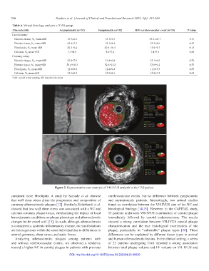
Figure 2. Representative case example of VH-IVUS analysis in the CAS patient.
contained more fibrolipids. A study by Samady et al. showed cerebrovascular events, but no difference between symptomatic
that wall shear stress alters the progression and composition of and asymptomatic patients. Interestingly, two animal studies
coronary atherosclerotic plaques [12]. Similarly, Eshtehardi et al. found no correlation between the VH-IVUS size of the NC and
showed that low wall shear stress was associated with a NC and histological findings [14,15]. However, in the CAPITAL study,
calcium coronary plaque tissue, emphasizing the impact of local 15 patients underwent VH-IVUS examination of carotid plaque
hemodynamic conditions on plaque phenotype and atherosclerotic immediately followed by carotid endarterectomy. The results
changes in the vessel wall [13]. As such, although atherosclerosis showed a strong correlation between VH-IVUS carotid plaque
is considered a systemic inflammatory disease, its manifestations characterization and the true histological examination of the
are heterogeneous within the same individual due to differences in plaque, particularly in “vulnerable” plaque types [16]. These
arterial geometry, shear stress, and static forces. differences can be explained by different tissue types in animal
Analyzing atherosclerotic plaques among patients with and human atherosclerotic lesions. In the clinical setting, a series
and without cerebrovascular events, we observed a tendency of 25 patients undergoing CAS reported a strong association
toward a higher NC in carotid plaques in patients with previous between total plaque volume and FF volume on VH IVUS and
DOI: http://dx.doi.org/10.18053/jctres.09.202304.23-00030