
Page 46 - JCTR-9-4
P. 46
262 Ali et al. | Journal of Clinical and Translational Research 2023; 9(4): 261-264
visit for elective cholecystectomy. She was referred to the medical
department for further workup after she was found to have
isolated elevated ALP levels of up to 1537 IU/L (reference range
40–150 IU/L). According to the patient she only had a history
of mild, non-radiating, non-pulsatile, bilateral headache that
occurred on and off several times during the day and that receded
without intervention. The patient had no history of deafness,
tinnitus, dental malocclusion, fractures, fatigue, or generalized
body weakness. She gave no associated history of any abnormal
bone enlargement, including her head, with no increase in hat size
in recent years.
On inquiring further, she reported a history of dark-colored
urine for the past 20 days with no history of pruritis, clay-colored
stool, or weight loss. Examination revealed mild frontal bossing,
whereas the rest of her general physical and systemic examination
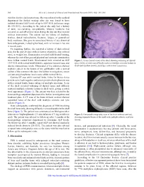
were within normal limits. Biochemical tests revealed an ALP Figure 1. X-ray (lateral view) of the skull showing widening of diploid
1537 IU/L with normal total bilirubin, aspartate transaminase, and space (white arrows) and diffusely scattered multiple sclerotic lesions in
alanine transaminase levels. Ultrasound of the abdomen showed skull vault (yellow arrow), yielding a cotton wool appearance.
multiple calculi in the lumen of the gallbladder with a normal
caliber of the common bile duct. On further investigations, serum
calcium and phosphorus levels were within normal limits.
Gamma GT was within normal limits. Urine for Bence–Jones
protein came back negative and serum protein electrophoresis was
within normal limits, hence ruling out multiple myeloma. X-ray
of the skull revealed widening of diploid space and diffusely
scattered multiple sclerotic lesions in skull vault, giving a cotton
wool appearance (Figure 1). The patient was then referred to the
rheumatology outpatient department for further investigations and
treatment plan. A CT scan of the brain without contrast showed
expanded bones of the skull with multiple sclerotic and lytic
lesions (Figure 2).
Bone scintigraphy confirmed the diagnosis of PDB involving
the skull bone only, showing diffusely increased uptake in the skull
bone (Figure 3). The patient was commenced on third-generation
intravenous bisphosphonate (15-min infusion of 5 mg zoledronic Figure 2. Computed tomography scan of the brain without contrast,
acid). The patient was advised to follow-up after 3 months at the showing expanded bones of the skull with multiple sclerotic and lytic
rheumatology outpatient department to determine ALP levels. lesions.
On follow-up after 3 months, repeat ALP test showed markedly
reduced levels down to 250 IU/L. The patient was also advised factors, and paramyxoviral infection [5]. Clinically, the usual
a repeat bone scintigraphy scan on the same visit but was lost to presentation is asymptomatic but may present with bone pains,
follow-up for subsequent visits. nerve entrapment, bony deformities, and increased propensity
to develop fractures. Clinical symptoms differ with the disease
3. Discussion
location; skull involvement usually causes increase in the hat size,
PDB is ranked second to osteoporosis as the most common along with headaches or deafness, whereas when base of the skull
bone disorder, exhibiting higher prevalence throughout Western is affected, it can lead to hydrocephalus, platybasia, and basilar
Europe, America, and Australia, but very low incidence among invagination [3,6]. High-output cardiac failure, although very
Asians and Africans. Diagnosis before the age of 40 is rare. The uncommon, is often precipitated due to enhanced vascularity of
key abnormality in this disease points to aberrant osteoclastic bone the affected bone, whereas osteosarcoma, although rare, is usually
resorption accompanied by marrow fibrosis, leading to increased the feared complication in a pre-existing pagetic bone [7].
bone vascularity and enhanced osteoblastic activity. A mosaic pattern According to the US Endocrine Society clinical practice
of bone and lamellar tissue with enlarged osteoclast-containing guidelines, effective management protocols have been formulated
distinct nuclear inclusion bodies is revealed histologically [4]. for patients suspected of PDB, where the first-line investigation
The etiology of PDB remains unclear, whereas multiple studies remains to be plain X-ray of the affected regions, followed
suggest an association with genetic mutations, environmental by 99m Tc-methylene diphosphonate (MDP) bone scan, after the
DOI: http://dx.doi.org/10.18053/jctres.09.202304.22-00186