
Page 68 - JCTR-9-4
P. 68
284 Al-Qahtani et al. | Journal of Clinical and Translational Research 2023; 9(4): 282-289
thrive (two males and six females). There was no significant Marsh–Oberhuber classification. Interestingly, most of these
association between gender and the different clinical presentations patients were females (74.6%), while only 18 were males.
(P = 0.97). The second most notable histopathological finding was an
Table 2 illustrates the histopathological findings of duodenal increased frequency of intraepithelial lymphocytes without villous
biopsies, grades of celiac disease according to Corazza and atrophy, which was found in 56 biopsies (37.3%) and classified as
Villanaci criteria, and histopathological classification according Grade A according to Corazza and Villanaci criteria, and Type 1/
to Marsh–Oberhuber criteria. Almost half of duodenal biopsies Type 2 lesions according to the Marsh–Oberhuber classification.
(71 cases, 47.3%) showed shortened villi caused by partial In 23 patients (15.3%), there was severe subtotal villous atrophy,
atrophy, which is consistent with Grade B1 according to Corazza assigned as Grade B2 according to Corazza and Villanaci
and Villanaci criteria, and Type 3A/3B lesions according to criteria and Type 3C lesions according to the Marsh–Oberhuber
classification. This group comprised 13 males (56.5%), compared
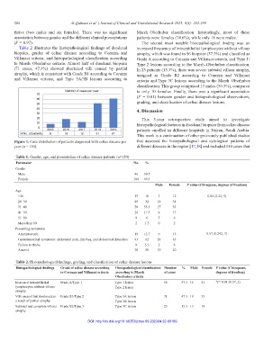
Number of cases per year to only 10 females. Finally, there was a significant association
70 (P = 0.01) between gender and histopathological observations,
60
50 grading, and classification of celiac disease lesions.
40 4. Discussion
30
20 This 5-year retrospective study aimed to investigate
10 histopathological features in duodenal biopsies from celiac disease
0
2015 2016 2017 2018 2019 patients enrolled in different hospitals in Najran, Saudi Arabia.
No. of patients 8 18 12 51 61 This work is a continuation of other previously published studies
Figure 1. Case distribution of patients diagnosed with celiac disease per that assessed the histopathological and cytological patterns of
year (n = 150). different diseases in the region [17,18] and included 150 cases that
Table 1. Gender, age, and presentation of celiac disease patients (n=150)
Parameter No. %
Gender
Male 46 30.7
Female 104 69.3
Male Female P value (Chi‑square, degrees of freedom)
Age
<20 15 10 3 12 0.82 (2.22, 5)
20–30 48 32 14 34
31–40 50 33.3 17 33
41–50 26 17.3 9 17
51–60 9 6 3 6
More than 60 2 1.3 0 2
Presenting symptoms
Asymptomatic 19 12.7 6 13 0.97 (0.242, 3)
Gastrointestinal symptoms: abdominal pain, diarrhea, and abdominal distention 93 62 28 65
Failure to thrive 8 5.3 2 6
Anemia 30 20 10 20
Table 2. Histopathological findings, grading, and classification of celiac disease lesions
Histopathological findings Grade of celiac disease according Histopathological classification Number % Male Female P value (Chi‑square,
to Corazza and Villanaci criteria according to Marsh– of cases degrees of freedom)
Oberhuber criteria
Increased intraepithelial Grade A/Type 1 Type 1 lesion 56 37.3 15 41 *P=0.01 (8.57, 2)
lymphocytes without villous Type 2 lesion
atrophy
Villi present but shortened as Grade B1/Type 2 Type 3A lesion 71 47.3 18 53
a result of partial atrophy Type 3B lesion
Subtotal and complete villous Grade B2/Type 3 Type 3C lesion 23 15.3 13 10
atrophy
DOI: http://dx.doi.org/10.18053/jctres.09.202304.22-00189