
Page 16 - MI-1-1
P. 16
Microbes & Immunity RANTES/CCL5 and ezrin peptide RepG3 for long COVID
Th2-type cytokines. RANTES/CCL5 also amplifies the
numbers of antibody-forming cells (AFCs) in both the
mucosa and systemically, resulting in increased antigen-
specific antibody titers both in mucosal secretions and in
the blood.
In mice, higher antigen-specific serum antibody titers
can be induced by nasal administration of antigen together
with RANTES/CCL5. For example, after three intranasal
doses of 100 µg of RANTES/CCL5 plus OVA antigen,
significantly higher serum titers of antigen-specific IgM
and IgG antibodies are produced relative to antigen
treatment alone. After RANTES/CCL5 plus antigen nasal/
mucosal administration, the most prevalent antigen-
specific antibody responses in the blood serum were
subclasses of IgG: IgG2a, IgG2b, IgG3, and IgG1, with later
elevation of IgG titers in the vaginal mucosa.
Soluble antigen plus RANTES/CCL5 induces the
proliferation of antigen-specific AFCs. Coadministration
of antigen plus RANTES/CCL5 significantly increased
antigen-specific IgA AFCs in the nasal cavity, upper and
lower respiratory tracts, Peyer’s patches, and intestine, and
also substantially increased antigen-specific IgM and IgG
AFCs in the spleen and respiratory tract lymphocytes but
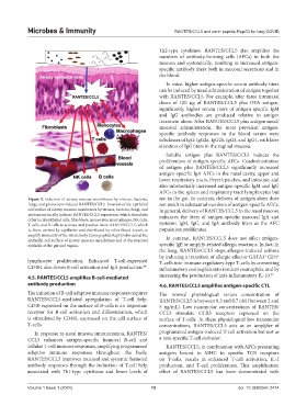
Figure 5. Infection of airway mucous membranes by viruses, bacteria, not in the gut. In contrast, delivery of antigen alone does
fungi, and protozoans induces RANTES/CCL5. Invasion of the epithelial not result in substantial numbers of antigen-specific AFCs.
cell surface of airway mucous membranes by viruses, bacteria, fungi, and In general, delivery of RANTES/CCL5 to the nasal mucosa
protozoans locally induces RANTES/CCL5 expression, which stimulates enhances the titers of antigen-specific mucosal IgA and
other local epithelial cells, fibroblasts, monocytes, macrophages, NK cells,
T-cells, and B-cells to activate and produce more RANTES/CCL5, which systemic IgM, IgG, and IgA antibody titers as the AFC
is, then, carried by capillaries and distributed by other blood vessels to population proliferates.
amplify immunity of the whole body. Ezrin peptide RepG3 also acts at the In contrast, RANTES/CCL5 does not affect antigen-
epithelial cell surface of airway mucous membranes and at the mucosal
epithelia of the gut and vagina. specific IgE or amplify-related allergic reactions. In fact, in
the lung, RANTES/CCL5 stops allergen-induced asthma
by inducing a transition of allergic effector GATA3 CD4
+
+
lymphocyte proliferation. Enhanced T-cell-expressed T-cells into immune-regulatory-type T-cells by converting
CD40L also drives B-cell activation and IgA production. 64 inflammatory eosinophils into resident eosinophils, and by
4.5. RANTES/CCL5 amplifies B-cell-mediated increasing the production of anti-inflammatory IL-10. 65
antibody production 4.6. RANTES/CCL5 amplifies antigen-specific CTL
The initiation of B-cell adaptive immune responses requires The normal physiological serum concentration of
RANTES/CCL5-mediated upregulation of T-cell help. RANTES/CCL5 is between 0.2 and 0.7 nM (between 2 and
CD40 expressed on the surface of B-cells is an important 5 ng/mL). Low nanomolar concentrations of RANTES/
receptor for B-cell activation and differentiation, which CCL5 stimulate CCR5 receptors expressed on the
is stimulated by CD40L expressed on the cell surface of surface of T-cells. At these physiological low nanomolar
T-cells. concentrations, RANTES/CCL5 acts as an amplifier of
In response to nasal mucosa immunization, RANTES/ programmed antigen-induced T-cell activation but not as
CCL5 enhances antigen-specific humoral B-cell and a non-specific T-cell activator.
cellular T-cell immune responses, amplifying programmed RANTES/CCL5, in combination with APCs presenting
adaptive immune responses throughout the body. antigens bound to MHC to specific TCR receptors
RANTES/CCL5 improves mucosal and systemic humoral on T-cells, results in enhanced T-cell activation, IL-2
antibody responses through the induction of T-cell help production, and T-cell proliferation. This amplification
associated with Th1-type cytokines and lower levels of effect of RANTES/CCL5 has been demonstrated with
Volume 1 Issue 1 (2024) 10 doi: 10.36922/mi.2474