
Page 41 - MI-1-2
P. 41
Microbes & Immunity Host receptors in immunogenic cell death
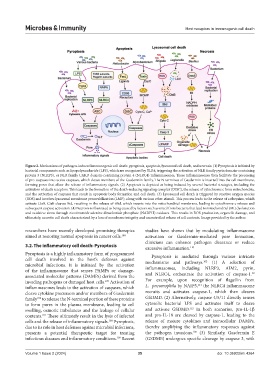
Figure 2. Mechanisms of pathogen-induced immunogenic cell death: pyroptosis, apoptosis, lysosomal cell death, and necrosis. (1) Pyroptosis is initiated by
bacterial components such as lipopolysaccharide (LPS), which are recognized by TLR4, triggering the activation of NLR family pyrin domain-containing
protein 3 (NLRP3), or NLR family CARD domain-containing protein 4 (NLRC4) inflammasomes. These inflammasomes then facilitate the processing
of pro-caspases into active caspases, which cleave members of the Gasdermin family. The N-terminus of Gasdermin is inserted into the cell membrane,
forming pores that allow the release of inflammatory signals. (2) Apoptosis is depicted as being induced by several bacterial strategies, including the
activation of death receptors. This leads to the formation of the death-inducing signaling complex (DISC), the release of cytochrome c from mitochondria,
and the activation of caspases that result in apoptotic body formation and cell death. (3) Lysosomal cell death is triggered by reactive oxygen species
(ROS) and involves lysosomal membrane permeabilization (LMP), along with various other stimuli. This process leads to the release of cathepsins, which
activate CtsB. CtsB cleaves Bid, resulting in the release of tBid, which inserts into the mitochondrial membrane, leading to cytochrome c release and
subsequent caspase activation. (4) Necrosis is illustrated as being caused by factors such as uracil from bacteria that lead to mitochondrial (Mt) dysfunction
and oxidative stress through nicotinamide adenine dinucleotide phosphate (NADPH) oxidases. This results in ROS production, organelle damage, and
ultimately, necrotic cell death characterized by a loss of membrane integrity and uncontrolled release of cell contents. Image provided by the author.
researchers have recently developed promising therapies studies have shown that by modulating inflammasome
aimed at restoring normal apoptosis in cancer cells. 104 activation or Gasdermin-mediated pore formation,
clinicians can enhance pathogen clearance or reduce
3.2. The inflammatory cell death: Pyroptosis
excessive inflammation. 110
Pyroptosis is a highly inflammatory form of programmed Pyroptosis is mediated through various intricate
cell death involved in the host’s defenses against 102
microbial infections. It is initiated by the activation mechanisms and pathways. (1) A selection of
of the inflammasome that senses PAMPs or damage- inflammasomes, including NLRP3, AIM2, pyrin,
39
associated molecular patterns (DAMPs) derived from the and NLRC4, orchestrate the activation of caspase-1.
105
invading pathogens or damaged host cells. Activation of For example, upon recognition of flagellin from
111
inflammasomes leads to the activation of caspases, which L. pneumophila by NAIP5, the NLRC4 inflammasome
cleave cytokine precursors and/or members of Gasdermin recruits and activates caspase-1, which then cleaves
family to release the N-terminal portion of these proteins GSDMD. (2) Alternatively, caspase 4/5/11 directly senses
106
to form pores in the plasma membrane, leading to cell cytosolic bacterial LPS and activates itself to cleave
112
swelling, osmotic imbalances and the leakage of cellular and activate GSDMD. In both scenarios, pro-IL-1β
contents. These ultimately result in the lysis of infected and pro-IL-18 are cleaved by caspase-1, leading to the
107
cells and the release of inflammatory signals. Pyroptosis, release of mature cytokines and intracellular DAMPs,
108
due to its role in host defenses against microbial infections, thereby amplifying the inflammatory responses against
presents a potential therapeutic target for treating the pathogen invasions. (3) Similarly, Gasdermin E
105
infectious diseases and inflammatory conditions. Recent (GSDME) undergoes specific cleavage by caspase 3, with
109
Volume 1 Issue 2 (2024) 35 doi: 10.36922/mi.4264