
Page 161 - MI-2-4
P. 161
Microbes & Immunity A novel anti-EphA8 monoclonal antibody
2.5. Determination of K by flow cytometry A
D
CHO/EphA8 and LN229/EphA8 were suspended
in 100 μL of serially diluted Ea Mab-9 (10 μg/mL to
8
0.0006 μg/mL), followed by treatment with Alexa Fluor
488-conjugated anti-mouse IgG (1:200). Fluorescence
data were subsequently collected using the BD FACSLyric
system (BD Biosciences, USA). The dissociation constant
(K ) was calculated by fitting the binding isotherms into
D
the built-in one-site binding model in GraphPad PRISM 6 B
(GraphPad Software Inc., USA).
2.6. Determination of K by enzyme-linked
D
immunosorbent assay (ELISA)
The recombinant EphA8-Fc (recEphA8) (Sino Biological
Inc., China) was immobilized on Nunc Maxisorp 96-well
immunoplates (Thermo Fisher Scientific Inc., USA) at
10 μg/mL for 30 min at 37°C. After washing with PBS containing C
0.05% Tween20 (PBST; Nacalai Tesque Inc., Japan), wells were
blocked with 1% BSA in PBST for 30 min at 37°C. The plates
were then incubated at serially diluted Ea Mab-9 (10 μg/mL
8
to 0.0006 μg/mL), followed by treatment with peroxidase-
conjugated anti-mouse IgG (1:2000; SouthernBiotech, USA).
1
Finally, enzymatic reactions were conducted using the ELISA
POD substrate TMB kit (Nacalai Tesque Inc., Japan). The K
D
value was determined as described above.
3. Results
3.1. Development of anti-EphA8 mAbs using the D
CBIS method
Currently, polyclonal antibodies against EphA8 for flow
cytometry are commercially available. However, they
are insufficient for therapeutic applications. Therefore,
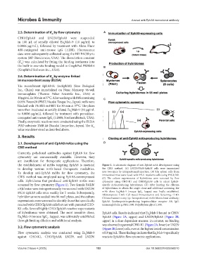
the establishment of mAbs targeting EphA8 is essential Figure 1. A schematic diagram of anti-EphA8 mAb development using
to develop various mAb-based therapeutic modalities. the CBIS method. (A) LN229/PA16-EphA8 cells were immunized
To develop anti-EphA8 mAbs for flow cytometry, the into two mice by intraperitoneal injection. (B) The spleen cells from
CBIS method was employed using EphA8-overexpressed immunized mice were fused with P3U1 myeloma cells using PEG1,500.
(C) The culture supernatants of hybridoma were screened by flow
cells. Hybridoma that produced anti-EphA8 mAbs were cytometry using CHO-K1 and CHO/EphA8 cells to select EphA8-
screened by flow cytometry (Figure 1). Two female BALB/ specific mAb-producing hybridomas. (D) After limiting the dilution
cAJcl mice were intraperitoneally immunized with LN229/ of hybridomas to obtain the single clone and additional screening, the
PA16-EphA8 cells once weekly for 5 weeks. Subsequently, mAb clone Ea Mab-9 (mouse IgG , kappa) was finally established.
8
1
hybridomas were seeded into 96-well plates, and the culture Abbreviations: CBIS: Cell-Based Immunization and Screening; CHO:
Chinese hamster ovary; i.p.: Intraperitoneal; mAb: Monoclonal antibody;
supernatants were screened to identify those that specifically EphA8: Erythropoietin-producing hepatocellular receptor A8; IgG:
reacted with CHO/EphA8 cells but not with parental CHO- immunoglobulin g; PEG1,500: Polyethylene glycol 1,500.
K1 cells. Several highly CHO/EphA8-reactive supernatants
of hybridomas were obtained. The most sensitive clone, EphA8 cells. Results indicated that Ea Mab-9 bound to CHO/
8
Ea Mab-9 (mouse IgG , kappa), was ultimately established EphA8 (Figure 2A, upper) and LN229/EphA8 (Figure 2B,
1
8
through limiting dilution and additional analysis. upper) in a dose-dependent manner. In contrast, no binding
was observed to parental CHO-K1 (Figure 2A, lower) or LN229
3.2. Flow cytometric analysis (Figure 2B, lower) cells, even at the highest tested concentration
Flow cytometric analysis was conducted using Ea Mab-9 of 10 μg/mL. These findings indicate that Ea Mab-9 specifically
8
8
against CHO-K1, CHO/EphA8, LN229, and LN229/ reacts to EphA8 in flow cytometric applications.
Volume 2 Issue 4 (2025) 153 doi: 10.36922/MI025060010