
Page 54 - MSAM-4-1
P. 54
Materials Science in Additive Manufacturing Additive manufacturing of 316L-Cu alloys
surface showed substantially reduced bacterial viability,
with CFU counts of 92 ± 25 and 114 ± 40 at 24 h and 48 h,
respectively, corresponding to 22% and 6% of the control
bacterial colonies, as shown in Figure 6B1 and B2.
A similar trend was observed on SS-5Cu surfaces, which
demonstrated even higher antibacterial efficacy, with
CFU counts of 63 ± 12 and 57 ± 10 at the same time
points, representing 15% and 3% bacterial viability,
respectively, as shown in Figure 6C1 and C2. While the
316L samples demonstrated a significant increase in CFU
between time points, the Cu-loaded samples effectively
suppressed bacterial growth, with minimal changes in
CFU counts from 24 h to 48 h. This inhibition of bacterial
proliferation highlights the role of Cu as an antimicrobial
agent.
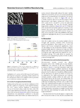
Figure 2. EDS mapping of the SS-5Cu surface revealed a homogenous
copper, chromium, and nickel distribution 4. Discussion
Abbreviation: EDS: Energy dispersive spectroscopy
316L SS is widely used in the medical industry for its
strength and corrosion resistance while maintaining
good biocompatibility. These properties are critical for
implant and fracture management applications, where
the environment of the human body presents a dynamic
situation ideally suited for corrosion and infection. When
alloyed with 316L, Cu has been shown to inhibit bacterial
proliferation and holds promising potential for use in
biomedical settings. This study aimed to measure the
mechanical and antibacterial properties of 316L-Cu alloys
produced through DED.
4.1. Microstructure and mechanical properties
Microstructural analysis revealed two distinct
formations within 316L. Cellular and columnar dendritic
solidification was observed in all three compositions,
Figure 3. XRD patterns of 316L, SS-3Cu, and SS-5Cu. Cu addition in as shown in Figure 1. Columnar formations result from
SS-3Cu and SS-5Cu resulted in an enlarged relative peak height at ~44°,
corresponding to face-centered cubic austenite the pronounced thermal gradient experienced during
Abbreviation: XRD: X-ray diffraction the DED process, with structures growing preferentially
along the heat flow direction towards the chilled substrate.
highlights an S. aureus cell exhibiting cell wall rupture On the other hand, the growth of cellular structures can
and exposed cytoplasm, providing visual evidence of be attributed to the rapid solidification rate, moderate
the antibacterial effect of Cu addition and its potential thermal gradients present within the melt pool, and
use in infection-resistant materials. lack of heat flow in the XY plane, all of which favor the
development of cellular dendritic structures. In contrast,
3.4. In vitro P. aeruginosa bacterial study traditional manufacturing methods, such as cold-rolling,
Antibacterial performance was further evaluated at 24 h begin with equiaxed austenitic grains and transform into
and 48 h against P. aeruginosa, Gram-negative bacteria. elongated martensitic structures due to the rolling process.
The 316L control surfaces displayed a pronounced Strain-induced martensite can be reversed into austenite
increase in bacterial colony count over time, with CFU through annealing, increasing its strength through grain
counts rising significantly from 422 ± 46 at 24 h to 1819 ± size reduction and achieving finer grain sizes than the
26
226 at 48 h, as seen in Figures 6A1 and A2. This increase original austenite.
demonstrates an environment conducive to bacterial The incorporation of Cu did not appear to significantly
proliferation on the 316L surface. In contrast, the SS-3Cu alter the microstructure of 316L, which is consistent with
Volume 4 Issue 1 (2025) 6 doi: 10.36922/msam.7357