
Page 18 - MSAM-4-2
P. 18
Materials Science in Additive Manufacturing Hydrogels in mandibular reconstruction
A
D
B
C
E
F
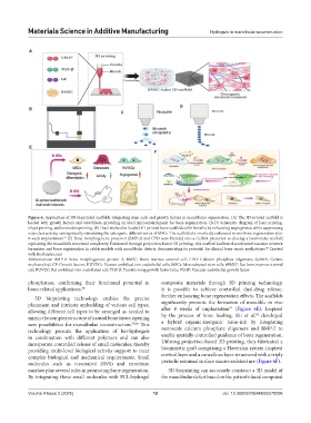
Figure 6. Application of 3D-bioprinted scaffolds integrating stem cells and growth factors in mandibular regeneration. (A) The 3D-printed scaffold is
loaded with growth factors and osteoblasts, providing an ideal microenvironment for bone regeneration. (B-D) Schematic diagram of laser printing,
inkjet printing, and extrusion printing. (E) Dual-molecules-loaded 3D-printed bone scaffolds offer benefits by enhancing angiogenesis while suppressing
osteoclast activity, synergistically stimulating the osteogenic differentiation of MSCs. The scaffold also markedly enhanced in vivo bone regeneration after
8-week implantation. (F) Bone morphogenetic protein-2 (BMP-2) and CPO were blended into a GelMA precursor to develop a biomimetic scaffold
81
replicating the mandible’s structural complexity. Fabricated through projection-based 3D printing, this scaffold facilitated accelerated vascular network
formation and bone regeneration in rabbit models with mandibular defects, demonstrating its potential for clinical bone repair applications. Created
82
with BioRender.com
Abbreviations: BMP-2: Bone morphogenetic protein 2; BMSC: Bone marrow stromal cell; CPO: Calcium phosphate oligomers; GelMA: Gelatin
methacryloyl; GF: Growth factors; HUVECs: Human umbilical vein endothelial cells; MSCs: Mesenchymal stem cells; rBMSC: Rat bone marrow stromal
cell; RUVEC: Rat umbilical vein endothelial cell; TGF-β: Transforming growth factor beta; VEGF: Vascular endothelial growth factor
phosphatase, confirming their functional potential in composite materials through 3D printing technology,
bone-related applications. 78 it is possible to achieve controlled dual-drug release,
3D bioprinting technology enables the precise further enhancing bone regeneration effects. The scaffolds
placement and intricate embedding of various cell types, significantly promote the formation of mandible in vivo
81
allowing different cell types to be arranged as needed to after 8 weeks of implantation (Figure 6E). Inspired
82
mimic the complex structure of natural bone tissue, opening by the process of bone healing, Shi et al. developed
new possibilities for mandibular reconstruction. 79,80 This a hybrid organic-inorganic nano-ink by integrating
technology permits the application of bio-hydrogels nanoscale calcium phosphate oligomers and BMP-2 to
in combination with different polymers and can also enable spatially controlled guidance of bone regeneration.
incorporate controlled release of small molecules, thereby Utilizing projection-based 3D printing, they fabricated a
providing multi-level biological activity support to meet biomimetic graft comprising a Haversian system-inspired
complex biological and mechanical requirements. Small cortical layer and a cancellous layer structured with a triply
molecules such as resveratrol (RVS) and strontium periodic minimal surface macro-architecture (Figure 6F).
ranelate play several roles in promoting bone regeneration. 3D bioprinting can accurately construct a 3D model of
By integrating these small molecules with PCL/hydrogel the mandibular defect based on the patient’s facial computed
Volume 4 Issue 2 (2025) 12 doi: 10.36922/MSAM025070006