
Page 19 - MSAM-4-2
P. 19
Materials Science in Additive Manufacturing Hydrogels in mandibular reconstruction
tomography (CT) or magnetic resonance imaging (MRI) osteoblasts and osteoclasts. Its molecular mechanisms
data, so that the hydrogel scaffold perfectly matches the shape involve coordinated regulation of multiple signaling
of the defect, achieving a highly personalized treatment pathways including Hypoxia-inducible factor 1-alpha
plan. This technique overcomes the difficulty of accurately (HIF-α), Wnt/β-catenin, Mitogen-activated protein kinases
replicating complex anatomical structures with traditional (MAPK), and PI3K/AKT/mTOR. These pathways not only
restorative materials. The unique rheological properties of regulate the directional differentiation of osteoprogenitor
the hydrogel, especially the shear thinning behavior, make it cells and functional expression of mature bone cells but
easy to flow when printing and quickly recover the viscosity also promote the balance between bone matrix anabolism
after deposition, ensuring high printing accuracy and and mineralization through activation of transcription
structural stability of the scaffold. In addition, 3D bioprinting factors such as RunX2 and Osterix.
technology optimizes the porosity and microstructure
of the scaffold by adjusting printing parameters such as 4.1. Hydrogels promote bone defect repair by
layer thickness and printing path, helping to promote regulating HIF-α signaling
cell migration and angiogenesis, thus providing an ideal HIF-α, a hypoxia-sensitive transcriptional regulator,
environment for bone tissue regeneration. Hydrogels can exhibits significantly upregulated expression levels under
also be loaded with growth factors or stem cells to provide hypoxic microenvironments. This pathway participates
bioactive support and speed up the repair process of the in bone development and remodeling through a dual
mandible. In mandibular defect models in rats and New regulatory mechanism. On the one hand, it transcriptionally
Zealand rabbits, 3D-printed scaffolds not only integrate activates pro-angiogenic factors such as VEGF, fibroblast
well with host tissues but also stimulated the growth of new growth factor 2, and platelet-derived growth factor to
bone tissue and vascular networks and successfully restored drive neovascular network formation (Figure 7A); on the
both the structural integrity and functional capacity of the other hand, it promotes osteogenic differentiation of MSCs
mandible. These results show the great potential of effluent and boosts bone matrix deposition. 86-89 The mechanism
gels in mandibular repair, representing a breakthrough by which the HIF-α signaling pathway facilitates bone
in the field of oral and maxillofacial surgery. The research growth is illustrated in Figure 7A. The activity of HIF-α
progress of 3D-bioprinted hydrogels used for the repair of is tightly regulated by cellular oxygen concentrations.
mandibular defects is summarized in Table 2. Hypoxic microenvironments created by specific
4. Mechanisms of hydrogels promoting hydrogel components such as deferoxamine (DFO) and
dimethyloxallyl glycine can effectively stimulate HIF-α
bone defect repair pathway activation. DFO, an iron-chelating compound
Bone defect repair is a complicated pathophysiological and hypoxia-inducing agent, upregulates VEGF expression
process that requires the dynamic balance between in hMSCs and human umbilical vein endothelial cells.
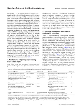
Table 2. Applications of 3D‑bioprinted hydrogels for the repair of mandibular defects
Composite hydrogels Cell type Animal model Outcome achieved References
3D-printed PCL/hydrogel composite MSCs, HUVECs Rat critical-sized Promoted mandibular bone formation after 81
with RVS and SrRn sustained releasing mandibular defect 8-week implantation
3D organic-inorganic nanoink with HUVECs Rabbit mandibular • Exceptional potential for osteogenesis and 82
BMP-2 and ultrasmall CPO incorporated defect angiogenesis in vitro
into GelMA precursor • Accelerated revascularization and
reconstructed neo-bone in vivo
3D-bioprinted multicellular GelMA/ Osteoblasts and In vitro Effective bioprinting of a mandibular 83
PEGDA scaffold endothelial cells structure
3D-printed bone frameworks of PCL/ MSC, EC In vitro • Effective blood vessel generation in vitro 84
HA and SVF-derived cell (SVFC) loaded and in vivo
bioink • Significant potential for craniofacial
skeletal defects
Coaxial 3D printing of HSM@HSA MC3T3-E1, BMSC Rat/Rabbit mandibular • Inhibited infection and inflammation 85
scaffold defect • Promoted osteogenesis and angiogenesis
Abbreviations: BMP-2: Bone morphogenetic protein 2; BMSC: Bone marrow stromal cell; CPO: Calcium phosphate oligomers; GelMA:
Gelatin methacryloyl; HA: Hydroxyapatite; HUVECs: Human umbilical vein endothelial cells; HSA: Hydroxyapatite-sodium alginate-antler
powders (HA-SA-APs); HSM: Hydroxyapatite-sodium alginate-minocycline hydrochloride (HA-SA-MINO); MSCs: Mesenchymal stem cells;
PCL: Polycaprolactone; PEGDA: Polyethylene glycol diacrylate; RVS: Resveratrol; SrRn: Strontium ranelate; SVF: Stromal vascular fraction.
Volume 4 Issue 2 (2025) 13 doi: 10.36922/MSAM025070006