
Page 78 - OR-1-1
P. 78
disease mechanisms and screen potential drugs in a more the accumulation of α-synuclein, the loss of dopaminergic
physiologically relevant environment compared to 2D neurons, and the activation of microglia. These organoids
149
cultures. 138,139,143,144 mimic the substantia nigra dopaminergic neurons, which
are essential for dopamine signaling to the basal ganglia.
5.2.2.1. MBO model They include not only midbrain dopaminergic neurons
Using chemically defined methods to differentiate expressing tyrosine hydroxylase (TH) but also astrocytes
midbrain floor plate neural progenitor cells (mfNPCs) and oligodendrocytes. These organoids are capable of
from pluripotent stem cells provides a crucial starting point forming complex neurotransmitter responses and possess
for generating midbrain-specific organoids (MBO). By the structure and function of autonomous neural networks.
145
applying key factors such as fibroblast growth factor 8 (Fgf8), Studies have shown that these mfNPCs can effectively
WNT signaling pathway, SHH, and dual SMAD inhibitors, differentiate into 2D midbrain dopaminergic neurons
researchers have successfully guided the differentiation of (mDAN) and 3D human midbrain-specific organoids
dopaminergic neurons. 146-148 (Figure 1). MBO models have (hMO), highly expressing markers associated with
captured key pathological features of the disease, including midbrain dopaminergic neurons, such as TH, FOXA2,
A B
C
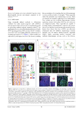
Figure 4. Application of organoids in Parkinson’s disease. (A) HP-β-CD stimulates neuronal differentiation and autophagy in patient-derived
organoids. HP-β-CD enhances neuronal differentiation by augmenting autophagy, as indicated by increased TFEB nuclear translocation, reduced p62
accumulation, and elevated LAMP1 and TH expression, with quantified protein expression dynamics and morphological assessments in organoids.
Reproduced with permission. Copyright © 2022 Wiley Periodicals LLC. (B) Temporal transcriptome analysis uncovers impaired development in
159
LRRK2-p. Gly2019Ser mutant midbrain organoids revealed developmental impairments through pseudotime analysis and gene expression patterns
compared to wild-type organoids. Reproduced with permission. Copyright © 2022 Elsevier Inc. (C) Integrative capacity of graft-derived dopaminergic
160
neurons in 6-OHDA-lesioned host mice. Human iPSC-derived midbrain organoids functionally integrate into the host’s neuronal circuitry, as evidenced
by immunohistochemical analysis of 6-OHDA-lesioned mice showing degeneration of endogenous TH-positive neurons and the successful innervation
of graft-derived DA neurons into the host substantia nigra and striatum. Copyright © 2023 Ivyspring International Publisher.
164
Abbreviations: HP-β-CD: 2-hydroxypropyl-β-cyclodextrin; iPSC: Induced pluripotent stem cell; LAMP1: Lysosomal associated membrane protein 1;
LRRK2: Leucine-rich repeat kinase 2; 6-OHDA: 6-hydroxydopamine; TFEB: Transcription factor EB; TH: Tyrosine hydroxylase.
Volume 1 Issue 1 (2025) 12 doi: 10.36922/or.8261