
Page 40 - OR-1-2
P. 40
A C
D
B
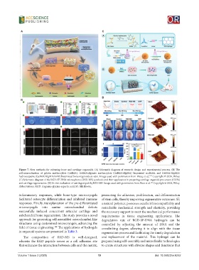
Figure 7. New methods for culturing bone and cartilage organoids. (A) Schematic diagram of research design and experimental process. (B) The
self-mineralization of gelatin methacrylate (GelMA), GelMA/alginate methacrylate (GelMA/AlgMA) bioprinted scaffolds, and GelMA/AlgMA/
hydroxyapatite (GelMA/AlgMA/HAP) bioprinted bone organoids in vitro. Image used with permission from Wang et al., Copyright © 2024, Wiley.
142
(C) Schematic diagram of the RGD-SF-DNA microspheres (RSD-MS) synthesis and their application in preparing cartilage organoid precursors (COPs)
and cartilage regeneration. (D) In vivo evaluation of cartilage repair by RSD-MS. Image used with permission from Shen et al. Copyright © 2024, Wiley.
143
Abbreviations: RGD: Arginine-glycine-aspartic acid; SF: Silk fibroin.
inflammatory responses, while bone-type microcryogels promoting the adhesion, proliferation, and differentiation
facilitated osteocyte differentiation and inhibited immune of stem cells, thereby improving regenerative outcomes. SF,
responses. Finally, transplantation of the pre-differentiated a natural polymer, possesses excellent biocompatibility and
microcryogels into canine osteochondral defects remarkable mechanical strength and elasticity, providing
successfully induced concurrent articular cartilage and the necessary support to meet the mechanical performance
subchondral bone regeneration. The study provides a novel requirements in tissue engineering applications. The
approach for generating self-assembled osteochondral-like degradation rate of RGD-SF-DNA hydrogels can be
structures using customized microcryogels, advancing the controlled by adjusting the amount of DNA and the
144
field of tissue engineering. The applications of hydrogels crosslinking degree, allowing it to align with the tissue
in organoid systems are presented in Table 3. regeneration process and facilitating the timely degradation
The composition of RSD-MS is well-designed, and replacement of the material. This hydrogel can be
wherein the RGD peptide serves as a cell adhesion site prepared using self-assembly and microfluidic technologies
that enhances the interaction between cells and the matrix, to create structures with diverse shapes and functions that
Volume 1 Issue 2 (2025) 19 doi: 10.36922/or.8262