
Page 66 - OR-1-3
P. 66
A B C D
E F G
H I
J
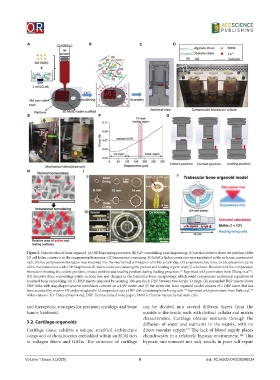
Figure 3. Construction of bone organoid. (A) 3D bioprinting processes; (B) Ca crosslinking post-bioprinting; (C) section views to show the position of the
2+
3D cell-laden construct in the compression bioreactor; (D) bioreactors containing 3D hMSCs-laden constructs were incubated in the in-house constructed
rack; (E) the compression bioreactor was mounted into the mechanical stimulation unit like a cartridge; (F) a representative force and displacement curve
of the mechanical test with a 3D bioprinted cell-laden constructs showing the preload and loading region; and (G) schematic illustration of the compression
bioreactor showing the culture position, contact position and loading position during loading processes. Reprinted with permission from Zhang et al. ;
131
131
(H) excessive bone remodeling results in bone loss and changes in the trabecular bone morphology, which could compromise anatomical regulation of
localized bone remodeling; (I) (i) DBP inserts prepared by securing 100-μm-thick DBP between two acrylic O-rings; (ii) suspended DBP inserts above
DBP disks with ring-shaped spacers; osteoblasts cultured on a DBP insert; and (J) the trabecular bone organoid model consists of a DBP insert that has
been activated by vitamin D3 and prostaglandin E2 suspended over a DBP disk containing bone lining cells. Reprinted with permission from Park et al. 132
132
Abbreviations: 3D: Three-dimensional; DBP: Demineralized bone paper; hMSCs: Human mesenchymal stem cells.
and therapeutic strategies for precision oncology and bone can be divided into several different layers from the
tumor treatment. outside to the inside, each with distinct cellular and matrix
characteristics. Cartilage obtains nutrients through the
3.2. Cartilage organoids diffusion of water and nutrients in the matrix, with no
141
Cartilage tissue exhibits a unique stratified architecture direct vascular supply. The lack of blood supply places
composed of chondrocytes embedded within an ECM rich chondrocytes in a relatively hypoxic environment. This
142
in collagen fibers and GAGs. The structure of cartilage hypoxic environment not only results in poor self-repair
Volume 1 Issue 3 (2025) 8 doi: 10.36922/OR025280024