Page 48 - TD-2-2
P. 48

Tumor Discovery                                                   Choroid plexus tumors: Benign to malignant


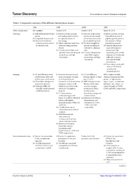
            Table 1. Comparative summary of the different choroid plexus tumors
                             CPA                 CPP                   aCPP               CPC
            WHO classification  Not classified   Grade I CPTs          Grade II CPTs      Grade III CPTs
            Histology        (i)  Well-differentiated tubular  (i)  Mitotic activity, necrosis,   (i)  Necrosis, brisk mitotic   (i) Mitotic activity, necrosis,

                               glands.             and nuclear pleomorphism   activity, and increased   high cellularity, loss of
                             (ii) Irregularly shaped acini.  are typically absent on   cell density and nuclear   papillary growth pattern,
                             (iii)  Basement membrane   microscopy.      pleomorphism present.  and invasion of brain
                                continuous and covered   (ii)  Bland columnar epithelium  (ii)  Portions of papillary   parenchyma present.
                                by cuboidal cells.  observed lining papillary   growth consisting of   (ii)  Grossly soft, globular
                                                   fronds.               cuboidal to columnar   mass with irregular
                                                 (iii)  Grossly pink, friable, soft,   cells.  projections, high
                                                    globular mass with irregular  (iii)  Grossly soft, globular   vascularity, and variable
                                                    projections, and high   mass with irregular   adherence to ventricular
                                                    vascularity.         projections, and high   walls with invasion
                                                                         vascularity.       into adjacent brain
                                                                                            parenchyma.
                                                                                          (iii)  Sectioning reveals solid
                                                                                             areas of intermixed
                                                                                             necrotic and
                                                                                             hemorrhagic foci.
            Imaging          (i)  CT and MRI may reveal   (i)  Prenatal ultrasound and   (i)  CT and MRI scan   MRI is highly variable,
                               calcified mass with solid   neurosonogram through   findings similar to those  demonstrating masses that
                               and/or cystic components.  an unfused anterior   seen in CPPs.  are iso- or hypointense,
                             (ii)  Ventricular enlargement   fontanelle will demonstrate  (ii)  MRI findings in more   hyper-, hypo-, or isointense,
                               may not be present.  echogenic lesions with   likely to show cysts,   and hypo-, iso-, hyper-, or
                             (iii)  Perfusion MRI will   bidirectional flow in the   necrotic changes,   mixed intensity on T1WI,
                                typically reveal increased   ventricles throughout   peritumoral edema,   T2WI, and DWI, respectively.
                                cerebral perfusion.  diastole.           unclear tumor
                                                 (ii)  CT demonstrates   boundaries, and greater
                                                   ventriculomegaly with   volume masses.
                                                   hyper- to isodense lobulated  (iii)  Masses are isointense,
                                                   lesions with slightly   hyperintense, and
                                                   irregular margins within the   isointense on T1WI,
                                                   ventricles.           T2WI, and DWI,
                                                 (iii)  Calcifications can be   respectively.
                                                    observed in about 25% of
                                                    patients.
                                                 (iv)  MRI demonstrates
                                                    flow voids and
                                                    enhanced well-defined
                                                    intraventricular lesions.
                                                 (v)  Masses are lobulated,
                                                   frond-like, and are
                                                   hypointense and
                                                   hyperintense on T1WI and
                                                   T2WI, respectively.
            Immunohistochemistry  (i)  Positive for cytokeratin,   (i)Positive for cytokeratin,   Positive for S-100,   (i)  Positive for cytokeratin,

                               S-100, transthyretin,   podoplanin, S-100, and   synuclein, and vimentin.  GFAP, S-100,
                               vimentin.           vimentin.                                transthyretin, and
                             (ii)  Variable expressivity   (ii)  Variable age-associated    vimentin.
                               of GFAP, CAM 5.2 and   expressivity of GFAP and            (ii)  GFAP positive in
                               cytokeratins 7 and 20.  transthyretin.                       roughly 20% of cases,
                                                                                            and positive staining for
                                                                                            S-100 and transthyretin
                                                                                            is typically less than
                                                                                            expected.
                                                                                          (iii)  May stain positive for
                                                                                             p53.
                                                                                                       (Cont’d...)


            Volume 2 Issue 2 (2023)                         6                          https://doi.org/10.36922/td.1057
   43   44   45   46   47   48   49   50   51   52   53