
Page 101 - TD-3-1
P. 101
Tumor Discovery PIN palsy due to lipoma
and lipoma are some of the non-neural sheath tumors
that induce compressive neuropathy, with lipoma being
regarded as the most common cause. Lipomas are benign
soft-tissue tumors arising from adipose tissue. They are
also called universal tumors because they can occur in
any part of the body . According to histopathological
[2]
findings, lipomas are divided into lipoblastoma (immature
fat cells), hibernomas (mature brown fat cells), and lipomas
(mature white fat cells). Lipomas can cause neuropathy in
four different ways: (i) Extrinsic compression by solitary
lipoma, (ii) intrinsic compression by encapsulated lipoma,
(iii) compression on the nerves by the fibrofatty masses
of lipofibromatous hamartomas, and (iv) compression by
macrodystrophia lipomatosa, causing localized gigantism
of the extremities, especially toes and fingers. Solitary
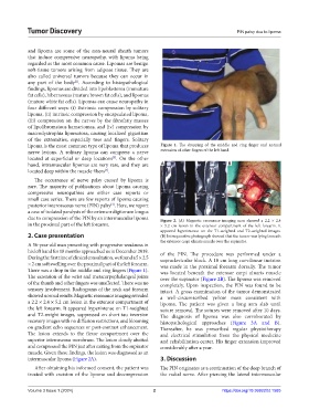
lipoma is the most common type of lipoma that produces Figure 1. The dropping of the middle and ring finger and normal
nerve lesions. A solitary lipoma can compress a nerve extension of other fingers of the left hand.
located at superficial or deep locations . On the other
[3]
hand, intramuscular lipomas are very rare, and they are A B
located deep within the muscle fibers .
[4]
The occurrence of nerve palsy caused by lipoma is
rare. The majority of publications about lipoma causing
compressive neuropathies are either case reports or
small case series. There are few reports of lipoma causing
posterior interosseous nerve (PIN) palsy . Here, we report
[4]
a case of isolated paralysis of the extensor digitorum longus
due to compression of the PIN by an intermuscular lipoma Figure 2. (A) Magnetic resonance imaging scan showed a 2.2 × 2.4
in the proximal part of the left forearm. × 5.2 cm lesion in the extensor compartment of the left forearm. It
appeared hyperintense on the T1-weighted and T2-weighted images.
2. Case presentation (B) Intraoperative photograph showed that the tumor was lying beneath
the extensor carpi ulnaris muscle over the supinator.
A 56-year-old man presenting with progressive weakness in
his left hand for 18 months approached us in December 2019. of the PIN. The procedure was performed under a
During the first time of clinical consultation, we found a 5 × 2.5 supraclavicular block. A 10 cm long curvilinear incision
× 2 cm soft swelling over the proximal part of the left forearm. was made in the proximal forearm dorsally. The tumor
There was a drop in the middle and ring fingers (Figure 1). was located beneath the extensor carpi ulnaris muscle
The extension of the wrist and metacarpophalangeal joints over the supinator (Figure 2B). The lipoma was removed
of the thumb and other fingers was unaffected. There was no completely. Upon inspection, the PIN was found to be
sensory involvement. Radiograms of the neck and forearm intact. A gross examination of the tumor demonstrated
showed normal results. Magnetic resonance imaging revealed a well-circumscribed yellow mass consistent with
a 2.2 × 2.4 × 5.2 cm lesion in the extensor compartment of lipoma. The patient was given a long arm slab until
the left forearm. It appeared hyperintense on T1-weighted suture removal. The sutures were removed after 10 days.
and T2-weight images, suppressed on short tau inversion The diagnosis of lipoma was also corroborated by
recovery images with no diffusion restrictions, and blooming histopathological approaches (Figure 3A and B).
on gradient echo sequences or post-contrast enhancement. Thereafter, he was prescribed regular physiotherapy
The lesion extends to the flexor compartment over the and electrical stimulation from the physical medicine
superior interosseous membrane. The lesion closely abutted and rehabilitation center. His finger extension improved
and compressed the PIN just after exiting from the supinator considerably after a year.
muscle. Given these findings, the lesion was diagnosed as an
intermuscular lipoma (Figure 2A). 3. Discussion
After obtaining his informed consent, the patient was The PIN originates as a continuation of the deep branch of
treated with excision of the lipoma and decompression the radial nerve. After piercing the lateral intermuscular
Volume 3 Issue 1 (2024) 2 https://doi.org/10.36922/td.1585