
Page 102 - TD-3-1
P. 102
Tumor Discovery PIN palsy due to lipoma
A The common causes for this entrapment are fracture
dislocation of the proximal radius and rheumatoid
arthritis. The milder form of radial tunnel syndrome
resembles the tennis elbow. The main structures causing
entrapment of the deep branch of the radial nerve in
the tunnel are the capsule-tendon-aponeurotic complex
in the anterior aspect of the radiohumeral joint, the
vascular arcade formed by the recurrent branch of the
B radial artery, and the arcade formed by the medial edge
of the extensor carpi radialis brevis and the arcade of
Frohse .
[7]
Compressive neuropathies of PIN may remain
undiagnosed for long. Sometimes, it may mimic tennis
elbow . The first case of parosteal lipoma surrounding the
[8]
neck of the radius causing weakness of finger extensors was
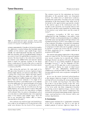
Figure 3. Hematoxylin-eosin-stained specimens showed mature reported by Richmond in 1953 . In an analysis of 31 cases
[9]
adipocytes without atypia and a uniform distribution of vacuoles in the
cytoplasm. (A) Low power ×10; (B) high power ×40. of non-traumatic PIN palsies, 14 patients had entrapment
of nerves within the supinator. The most common cause
septum at approximately 10 cm above the lateral epicondyle, of compression in their series was the ganglion, followed
[10]
the radial nerve traverses between the brachialis muscle by lipoma . Another case showed that the excision of
medially and the extensor carpi radialis longus muscle soft-tissue chondroma led to a complete resolution of
[11]
laterally. The lateral epicondyle branches into superficial symptoms of PIN palsy .
and deep branches, and the superficial branch continues The anterior and posterior approaches to the proximal
as a sensory branch through the flexor compartment of the radius can injure the PIN. The anterior approach requires
forearm. The deep branch of the radial nerve innervates more muscle retraction but is relatively safer. Besides,
the extensor carpi radialis brevis and supinator muscle Monteggia fracture can produce PIN palsy either at the
before it enters the supinator between its two heads. time of injury or occur during treatment of neglected
Then, it winds around the neck of the radius to reach the Monteggia fractures [12,13] . van den Bogaerde and Shin
posterior compartment of the forearm. From here onward, reported a case of PIN palsy due to nerve incarceration
it is known as the PIN. with EndoButton during the repair of distal biceps
[14]
After exiting the supinator, the main trunk of the rupture . Of note, inflammatory myofibroblastic
PIN gives three branches to extensor digitorum longus, pseudotumors, rheumatoid arthritis, false aneurysm, and
extensor digiti minimi, and extensor carpi ulnaris. Then, psoriatic arthritis rarely cause compressive neuropathy of
it divides into a long branch which innervates extensor the PIN [15-18] .
pollicic longus and extensor indicis and a short branch In our case, the patient developed isolated paralysis
innervating abductor pollicic longus and extensor pollicis of the extensor digitorum longus of the left forearm
brevis. There are many variations in the origin of the and had difficulty extending the metacarpophalangeal
branch to the extensor carpi radialis brevis. It can originate joint of the middle and ring fingers. We believe that
from the radial nerve, the superficial branch of the radial this is due to the peculiar anatomy of the PIN. After
nerve, or the PIN . The clinical manifestations of PIN winding around the neck of the radius, the main trunk
[5]
palsy vary according to the site of involvement. The long of the nerve splits into three independent branches
radial extensors are usually spared in typical PIN palsy. innervating extensor digitorum longus, extensor digiti
Usually, the ulnar extensor and long extensor of fingers minimi, and extensor carpi ulnaris. Compression only
will be affected. When the patient is attempting to extend occurred to the branch innervating extensor digitorum
the wrist, there is a radial deviation due to the unopposed longus, sparing branches to the extensor indicis, and
action of the extensor carpi radialis longus. There will extensor digiti minimi.
not be any associated deep tendon reflex or sensory
abnormalities in PIN palsy . 4. Conclusion
[6]
PIN syndrome may result in finger and thumb drops. Isolated muscle weakness due to compressive neuropathy
This is due to the entrapment of the deep branch of the of the PIN is extremely rare. This is a rare case of
radial nerve between the two heads of the supinator. isolated paralysis of extensor digitorum longus due to
Volume 3 Issue 1 (2024) 3 https://doi.org/10.36922/td.1585