
Page 107 - TD-3-1
P. 107
Tumor Discovery Odontogenic myxofibroma arising in a child with long-term follow-up
A B C
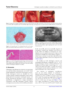
Figure 4. Intraoperative findings. (A) Surgical exposure of the lesion. (B) The lesion after tumorectomy. (C) The lesion after iliac cancellous bone grafting.
An absorbable plate was applied to give the mandibular angle morphology, and the gap between the plate and defect was filled with iliac cancellous bone.
Figure 7. A panoramic X-ray scan at 8-year postoperatively. The grafted
bone at the right mandibular angle had been integrated with the existing
bone, and the mandibular angle morphology was found to be under
Figure 5. Enucleated specimen. The enucleated specimen, with a glossy recovery (arrows).
surface and translucent white color, measures 17 × 15 × 10 mm in size.
in the mucous-like matrix are specifically diagnosed as
A B odontogenic myxofibromas. Typically, lesions that occur
in the jawbone are defined as odontogenic, but such a
definition does not extend to the presence or absence of an
odontogenic epithelial mass while making a diagnosis. 1,4,10
Indeed, many cases have actually been reported in which
no odontogenic epithelium is found, as in our present case
(Tables 1 and 2).
Figure 6. (A and B) Histopathological findings (hematoxylin-eosin The incidence of both odontogenic myxoma and
stain). Both the image at different magnification showed. spindle-shaped odontogenic myxofibroma is estimated to be 3.3 – 15.7%
cells with little atypia had proliferated in the mucous-like matrix, and the among odontogenic tumors in Europe and the US. The
5,7
presence of odontogenic epithelium was unclear.
incidence in children under 10 years of age is 0.7 – 3.5%,
which is lower than that in adults. In Japan, the incidence
1-3
3. Discussion of odontogenic myxoma and myxofribroma is 1.6 – 2.7%,
Odontogenic myxofibromas are relatively rare tumors that which is lower than that in other countries.
occur in the jawbone intraosseously. In the 2017 World Since statistics on odontogenic myxofibromas
Health Organization classification of odontogenic tumors, alone are not widely available, we performed a
9
benign odontogenic tumors are classified into three clinical statistical analysis of 46 cases of odontogenic
categories: Benign epithelial odontogenic tumors, benign myxofibromas of the mandible occurring in Japan from
mixed epithelial-mesenchymal odontogenic tumors, and 1970 to 2023. The patient age ranged from 3 to 64 years
benign mesenchymal odontogenic tumors. Odontogenic old, and 38 cases (82.6%) were ≥11 years old (Table 1),
myxofibroma is categorized as a benign mesenchymal while 8 (17.4%) were ≤10 years old (Table 2). There
odontogenic tumor, like odontogenic myxoma. Among was a slight preponderance of female patients in the
odontogenic myxomas, those with a lot of fibrous formation cases, with 17 male cases (44.7%) and 21 female cases
Volume 3 Issue 1 (2024) 3 https://doi.org/10.36922/td.2096