
Page 106 - TD-3-1
P. 106
Tumor Discovery Odontogenic myxofibroma arising in a child with long-term follow-up
We herein report a pediatric case of odontogenic signal intensity on T2-weighted short inversion time
myxofibroma confined to the mandibular angle, who had inversion-recovery imaging with a partially irregular
experienced an uneventful postoperative course with no border with the surrounding bone (Figure 3). Because
recurrence after more than 14 years of follow-up. the possibility of a malignant tumor could not be ruled
2. Case presentation out merely based on the imaging findings, a biopsy was
performed, and a histological evaluation of the biopsied
In June 2009, a 10-year-old Japanese boy presented to our sample rule out a diagnosis of osteosarcoma in our
department following a complaint of a painless swelling patient. The histopathological findings demonstrated
in the right mandibular angle. The pediatric patient had fibrous proliferation with bland spindle cells, and the
no remarkable personal or family medical history. Clinical immunostaining showed negative expression of AE1/AE2
findings included a palpable bone-like hard swelling in and desmin, and positive expression of SMA and bcl2. The
the right mandibular angle, but there were no abnormal diagnosis was deemed to be odontogenic myxofibroma or
findings in the oral cavity. chondromyxofibroma.
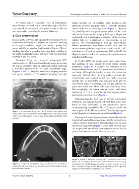
Panoramic X-ray and computed tomography (CT) In October 2009, the patient underwent tumorectomy
scan revealed an ill-defined radiolucent image measuring and grafting of iliac cancellous bone under general
20 mm in diameter with an indistinct border, featuring anesthesia (Figure 4). A surgery was operated on the
a bone-like protrusion in the right mandibular angle submandibular lesion, and the tumor was enucleated in
(Figures 1 and 2). Magnetic resonance imaging revealed one lump. Although the border between the tumor and
low signal intensity on T1-weighted imaging and high bone was relatively clear, the bony surface exposed after
tumorectomy was curetted in one layer with a rounded
carbide bur. An absorbable plate was applied to give the
mandibular angle morphology, and the gap between the
plate and defect was filled with iliac cancellous bone.
Macroscopically, the tumor was an elastic, soft lesion
measuring 17 × 15 × 10 mm in size with a glossy surface
and translucent white color (Figure 5).
Histopathologically, there was no obvious odontogenic
epithelium, and spindle-shaped cells with little atypia were
found to have proliferated in the mucous-like matrix.
Immunostaining showed that AE1/AE2, desmin, and S-100
Figure 1. A panoramic X-ray scan. An ill-defined bone defect with were negative, while SMA was positive. These findings support
cortical bone resorption and the outward protrusion of the surrounding a final diagnosis of odontogenic myxofibroma (Figure 6).
cortical bone-like spicules could be seen in the right mandibular angle Panoramic X-ray at 8 years postoperatively showed that
(arrows).
the grafted bone had been integrated with the residual bone,
without obvious difference in mandibular growth between
the left and right sides (Figure 7). More than 14 years since
the surgery had passed, the post-operative course for the
patient had been uneventful without recurrence.
A B
Figure 3. Magnetic resonance imaging. (A) T1-weighted image (axial
plane). (B) T2-weighted short inversion time inversion-recovery image
Figure 2. Computed tomography image obtained in the axial plane. (axial plane). T1-weighted image showed a low signal intensity (arrows),
A bone-like spicule was observed around the cortical bone defect with an and T2-weighted image showed a high signal intensity with a partially
unclear border (arrows). irregular border with the surrounding bone (arrows).
Volume 3 Issue 1 (2024) 2 https://doi.org/10.36922/td.2096