
Page 112 - TD-3-3
P. 112
Tumor Discovery Columella fistula
seizures. He was referred to us from the pediatric 3. Discussion
department, where he was being followed due to the
observation by his pediatrician of a fistula at the level Cysts and fistulas of the dorsum of the nose are rare
malformations, strictly confined to the midline, extending
of the columella, with pus discharge in the context of from the columella to the glabella. Occasionally,
2-4
a dysmorphic syndrome (Figure 1). After conducting one or more hairs are present, centered on the fistula.
the clinical examination, we requested a brain scan A deep extension is possible, potentially contacting
to investigate any communication between the fistula the leptomeningeal spaces. Pre-operative imaging is
and the base of the skull. The first reading of the scan systematically carried out to determine the depth of
suggested a suspicion of communication with the anterior extension and the relationship of the malformation with
level of the base of the skull, which was ruled out after the cribriform lamina and meninges. Surgical excision
magnetic resonance imaging (MRI) showed that the is preferably performed before the age of one due to the
columella fistula was superficial (Figure 2). The patient risk of infection; neurosurgical management is required if
was then scheduled for excision of the fistula. The surgical communication with the meningeal spaces is suspected. 4-6
procedure involved making an incision next to the cyst
at the level of the columella, followed by step-by-step Among the various pathological theories proposed, the
dissection until reaching the insertion of the cord at the most widely accepted is that proposed by Grunwal in 1910
7-9
level of the nasal bones and reaming along the line uniting and later developed by Pratt.
the nasal bones. Embryologically, between the frontal and nasal bones
lies a membrane called the fonticulus nasofrontalis.
Behind it, an extension of the dura mater insinuates itself.
During development, this extension is enveloped by a
bony canal whose entrance is called the foramen cecum,
which eventually closes completely with growth. When
this closure does not occur, herniations of the cerebral
envelopes can happen, sometimes accompanied by
cerebral parenchyma. 10,11 In such cases, the development of
mesoblastic tissue halts, leaving remnants of nervous tissue
that may remain in the same region, giving rise to gliomas
or ectodermal tissue forming dermoid cysts. 11,12
Gliomas, dermoid cysts, and meningoencephaloceles
are the differential diagnoses to consider in the presence
of a medial frontal malformation or a malformation of the
dorsum of the nose. Nasal gliomas are rare congenital tumors
composed of heterotopic neuroglial tissue. Due to the risk of
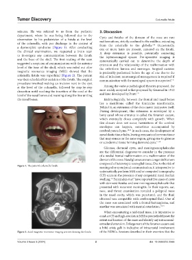
Figure 1. The patient’s columella fistula
meningeal or ependymal communication, it is imperative to
systematically perform MRI and/or computed tomography
(CT) scans in the presence of any congenital nasal median
swelling. Fernàndez et al. have reported five cases of cysts
3,4
3
with dermoid fistulas and one meningoencephalocele that
presented with recurrent meningitis. In their reports, ear,
nose, and throat examination revealed a polypoid mass
in the nasal cavity, which was punctured, and the fluid
obtained was compatible with cerebrospinal fluid. One of
the cases was associated with a frontal hemangioma, and
another was associated with mental retardation. 12,13
When encountering a mid-nasal mass, it is imperative to
conduct CT and high-resolution MRI to precisely delineate the
extent and location of the mass and identify any intracranial-
extradural extension. Enlargement of the foramen cecum and
a bifid crista galli is indicative of intracranial involvement
Figure 2. Axial magnetic resonance imaging section showing the fistula of the NDSCs. Sessions described in their overview that the
Volume 3 Issue 3 (2024) 2 doi: 10.36922/td.2566