
Page 170 - TD-3-4
P. 170
Tumor Discovery Signet ring cell carcinoma of the urinary bladder
Adenocarcinomas are the most prevalent histologic A B
type that manifests as CUP. SRCC, a poorly differentiated
aggressive subtype of adenocarcinoma, has been rarely
reported to manifest as metastatic SRCC of unknown
primary origin. This case report describes a patient who
11
presented with skeletal metastasis and circulating tumor
cells (CTCs) exhibiting signet ring (SR) morphology. The
diagnosis of SRCC was confirmed through bone marrow
biopsy results. Despite an in-depth diagnostic workup, we
were unable to establish the primary site of the tumor. The
patient died 2 months after hospitalization. At autopsy, the
urinary bladder was established as the primary site of the
tumor.
2. Case presentation
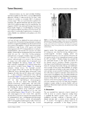
A 65-year-old man was admitted for severe asthenia and Figure 1. Findings of skeletal involvement (A) Tc-99m-methylene-
diphosphonate scintigraphy revealing a super-scan pattern with multiple
low-grade fever. He reported having previously undergone foci of increased tracer uptake throughout the skeleton, with little or no
mandibular surgery for ameloblastoma. Blood tests revealed activity in the soft tissues or urinary tract. (B) Computed tomography
severe anemia (Hemoglobin = 3.8 g/dL), thrombocytopenia (sagittal plane) image revealing mixed (lytic and sclerotic) lesions in the
(34.000/mm ), high ferritin (2100 ng/mL) and lactate vertebral bodies.
3
dehydrogenase levels (777 U/L), hypokalemia (1.61 mEq/L),
hypoproteinemia, and normal liver enzymes and tumor negative results. Two peripheral lesions indeterminate
markers. Serum alkaline phosphatase levels were elevated for malignancy were identified during multiparametric
(1102 U/L, n.v. 34 – 102 U/L). Tc-99m-methylene- prostate-magnetic resonance imaging, which was
diphosphonate scintigraphy revealed a super-scan pattern conducted in response to the low free/total PSA ratio and
(i.e., there was a concentration of the radiotracer in the the negative results of the immunohistochemical staining
skeleton with minimal or no activity in the soft tissues for CK7 and CK20. 1,3-6 Prostatic biopsy was planned but
or urinary tract) with multiple foci of increased tracer not performed because of the rapid deterioration of
uptake throughout the skeleton (Figure 1A). Whole- the patient’s general conditions (Eastern Cooperative
body computed tomography (CT) revealed mixed (lytic Oncology Group/World Health Organization Performance
and sclerotic) vertebral lesions (Figure 1B), indicating Status 4). Androgen deprivation therapy and zoledronate
metastatic skeletal disease. The free/total prostate- were initiated, but the patient died suddenly. An autopsy
specific antigen (PSA) ratio was 9.41. The FDA-approved revealed pulmonary embolism as the cause of death. The
CellSearch system (Menarini Silicon Biosystems, Castel entire prostate was processed for histological examination
®
Maggiore, Bo, Italy) detected 387 solitary and 9 clustered due to the clinical suspicion of prostate cancer; however, it
(2 – 3 cells) cytokeratin (CK)-positive CTCs with SR was revealed to be malignancy-free. The urinary bladder was
morphology (Figure 2A), as previously described. Briefly, devoid of any intraluminal mass. Histological examination
12
7.5 mL of whole blood was processed using the CellSearch revealed diffuse SRCC cell infiltration within the entire
®
CTC kit. After EpCAM-based immunomagnetic thickness of the lamina propria (Figure 2D and E). Rare
capture, the cells were stained with antibodies anti- cells were immunoreactive for CK7 and CK20. Neoplastic
CK8,18,19-fluorescein isothiocyanate and anti-CD45- lymphatic invasion within the urinary bladder wall was
allophycocyanin and with 4′,6-diamidino-2-phenylindole significant. Mesenteric lymph nodes had metastases along
for detecting the nucleus. Immunofluorescence images the vertebrae.
®
were eventually analyzed using CellSearch Analyzer 3. Discussion
II. SRCC metastasis was diagnosed on the iliac crest
and vertebral biopsies (Figure 2B and C). The neoplastic The case reported here represents a typical example of
cells tested negative for CK7, CK20, TTF1, and PSA. CUP, in which an extensive diagnostic workup failed
Because SRCC is known to originate more frequently from to establish the tumor’s primary origin. CUP, a well-
the gastrointestinal tract, esophagogastroduodenoscopy, established oncologic condition, is associated with a
colonoscopy, small-intestine contrast ultrasonography, dismal outcome and a median survival duration of
and CT enterography revealed negative results. Transrectal 8 – 11 months. Its diagnosis is always based on the
1-4
ultrasonography and urinary cytology also revealed histopathologic examination of the biopsy specimen
Volume 3 Issue 4 (2024) 2 doi: 10.36922/td.3736